Dr. Phil Zeltzman’s Blog
Squirrel gets a new lease on life after cleft palate surgery
Squirrel, a 2 year old bully mix, had an unusual problem.
She was struggling with eating and drinking on her own, as well as playing and chewing on toys.
Thick, white mucus would build up and make it hard for her to swallow or breathe through her nose.
Because of that, it was difficult for her to gain weight.
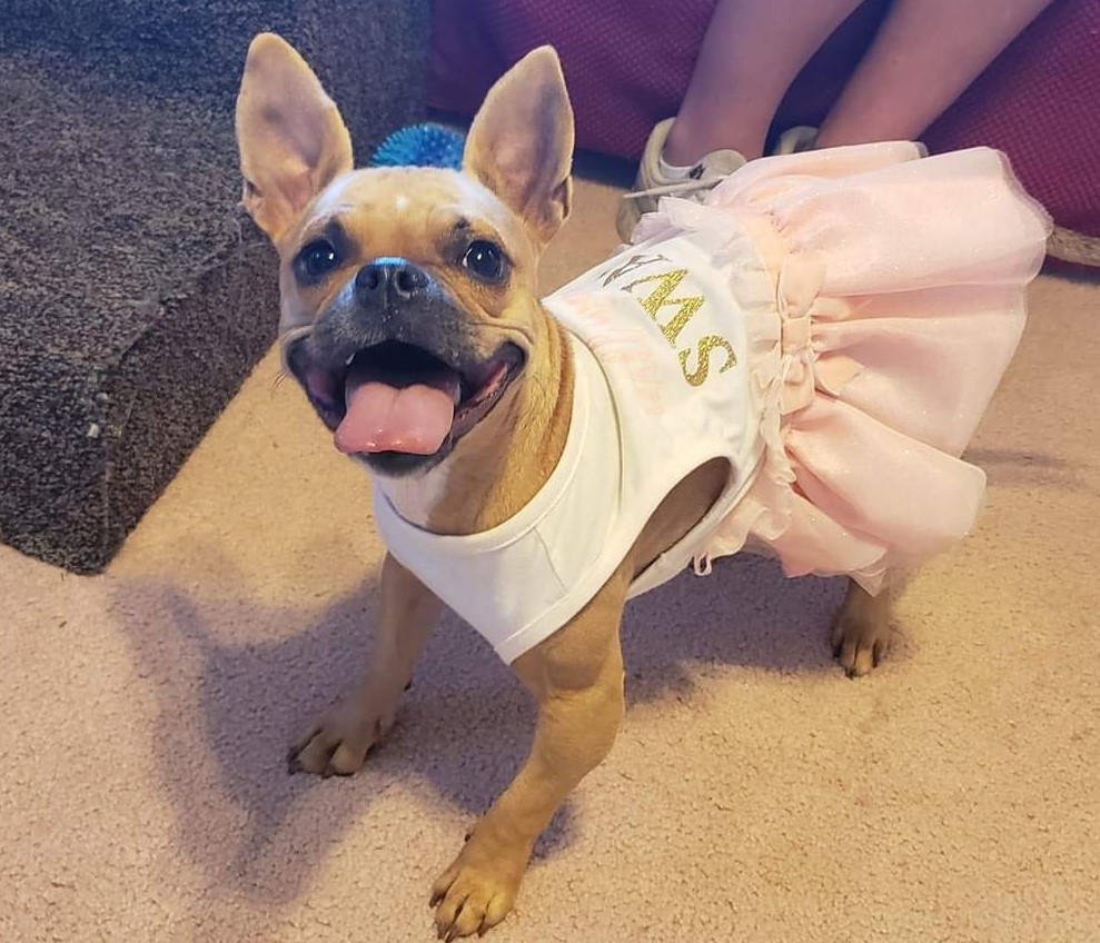
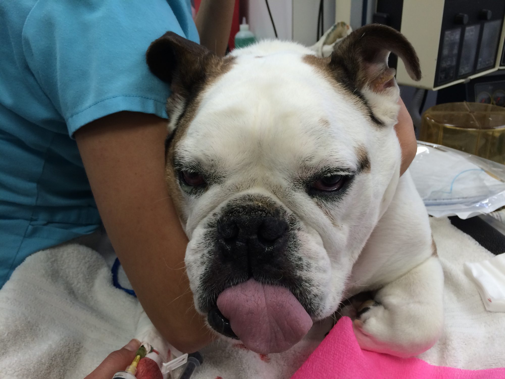
This is how she looked when she was first adopted.
Her foster explained: “She is choking & sneezing food and mucus out her nose every day, anytime she moves around, plays or chews on toys.”
“We almost lost her a few times the first year,” said her foster. “So she is a fighter for sure. She is a spunky little thing loving life!”
Her diagnosis? A cleft palate.
This means that there was a split in the roof of the mouth (similarly, a cleft lip is a split in the lip).
That split allowed food and water to go up in the nasal passages, causing difficulty breathing and constant infections.
That also meant that she could not get the proper nutrition her body needed to grow properly.
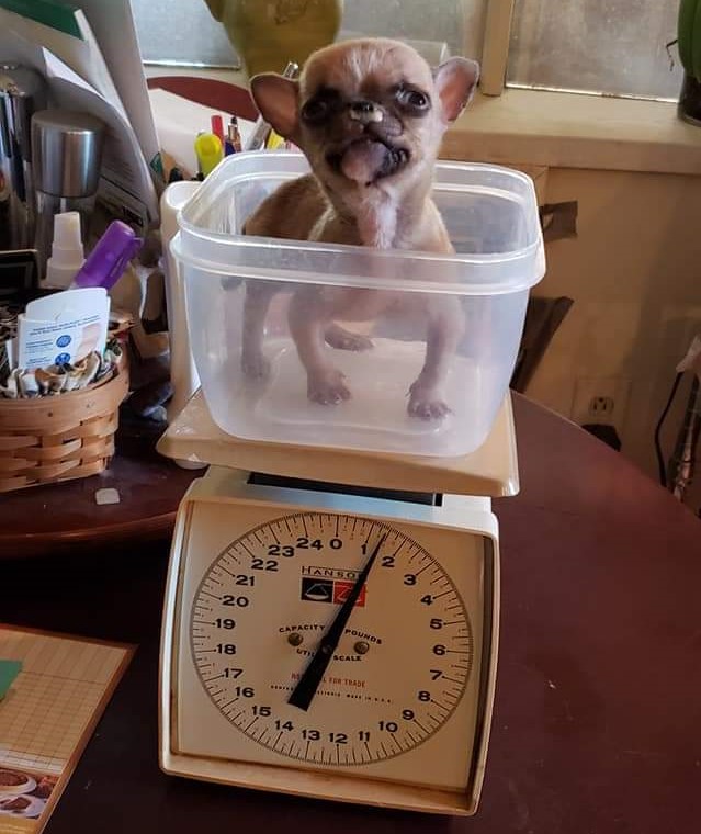
This is the reason why a puppy (or a kitten) with a cleft palate is typically the runt of the litter.
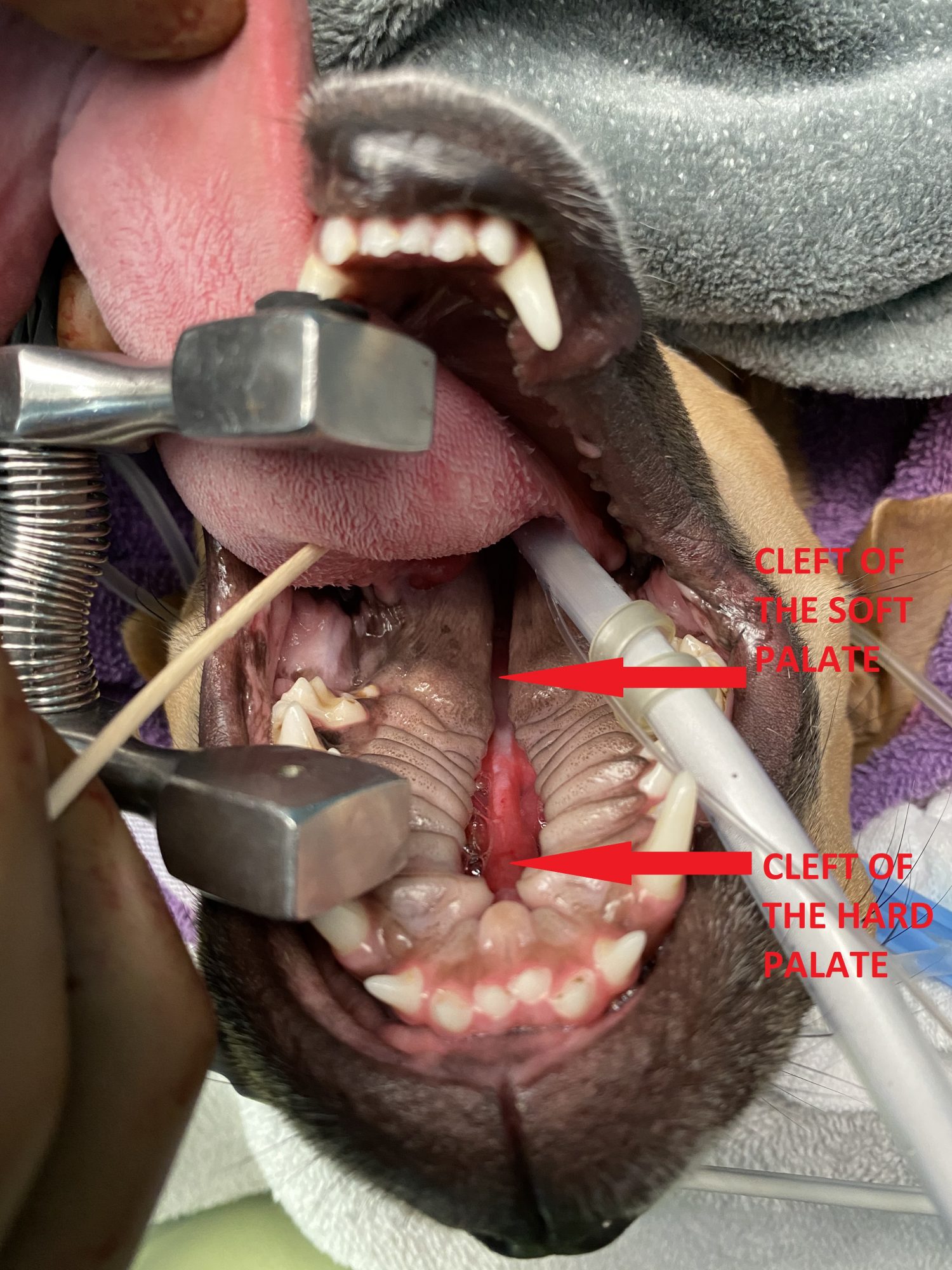
Now, a bit of anatomy. When you touch the roof of your mouth, it is firm because there is bone behind it. You are touching your “hard” palate.
PICTURES OF THE CLEFT BELOW ARE GRAPHIC AND ARE NOT FOR THE FAINT OF HEART.
The bone then disappears in the back of the mouth, so that part of the palate is called the “soft” palate.
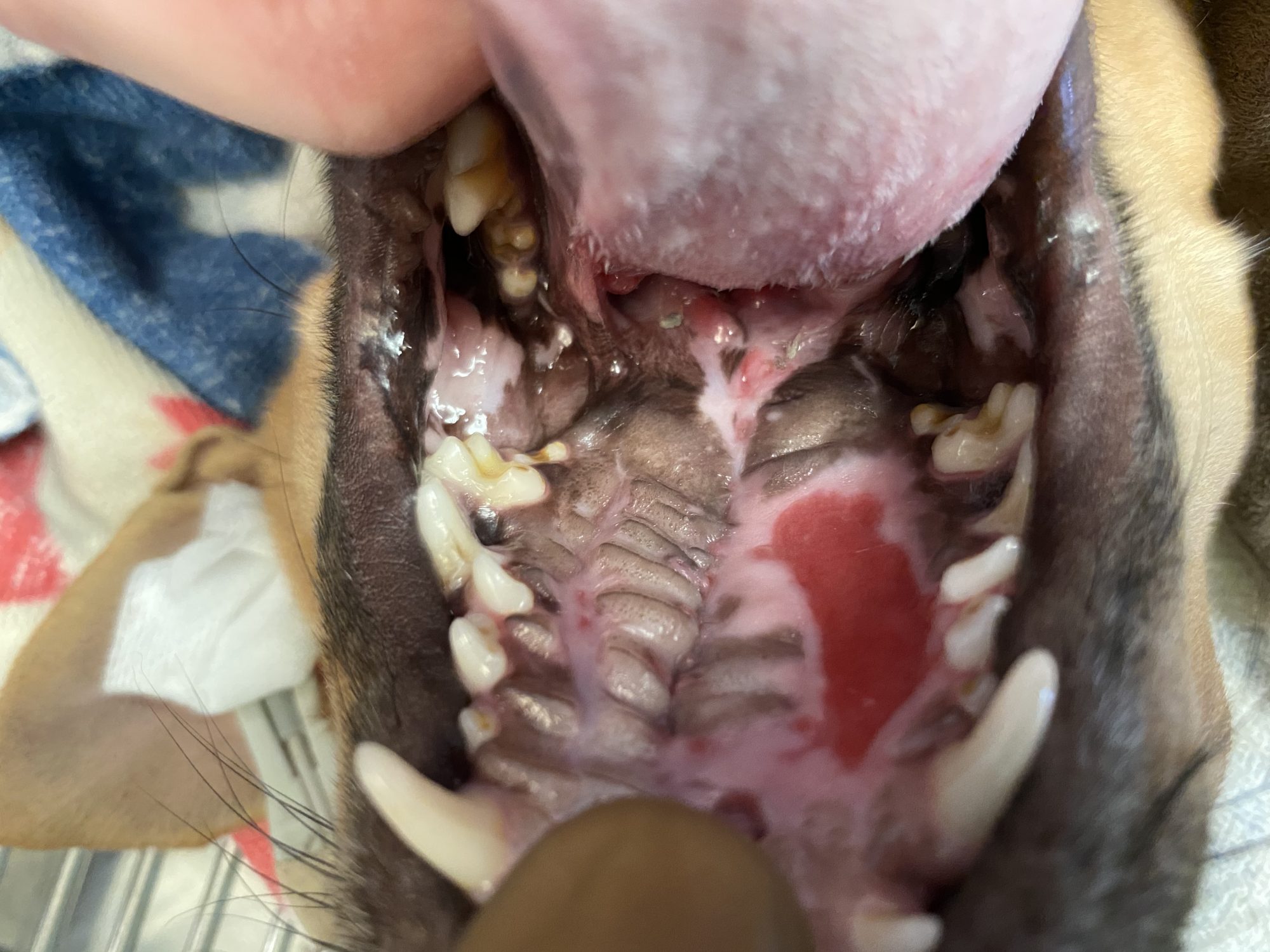
Based on preop pictures, Squirrel clearly had a cleft of the hard palate.
The surprise came when we put her under anesthesia. A more thorough exam revealed a cleft in 100% of the roof of the mouth. So little Squirrel had a cleft of the hard palate and the soft palate.
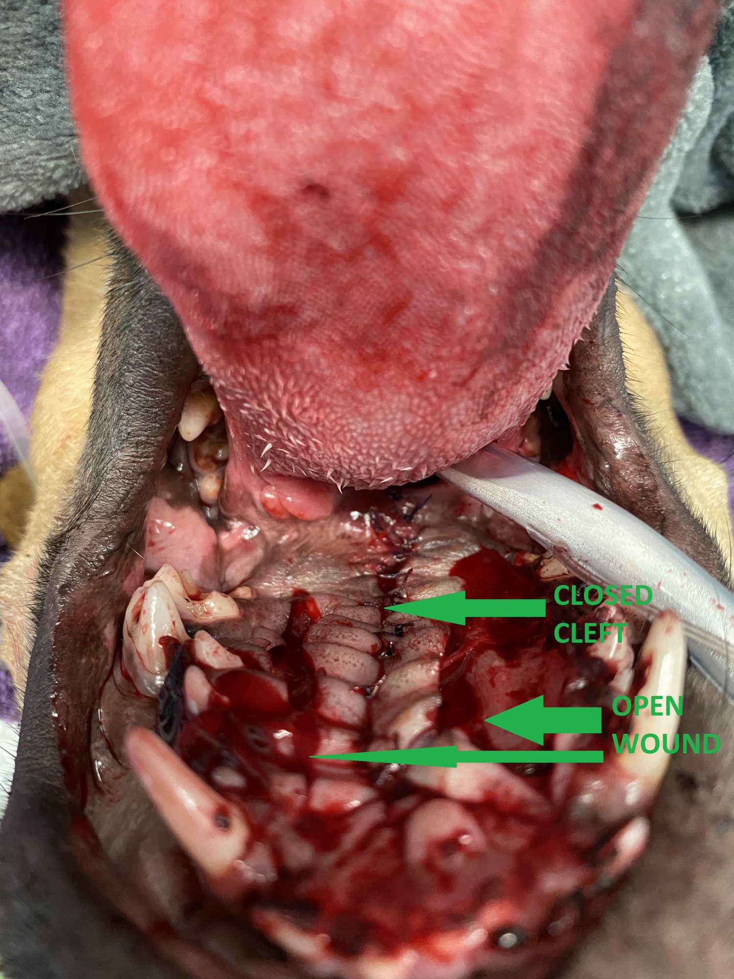
In all of the pictures of the mouth below, Squirrel is on her back, and you see the inside of the roof of the mouth.
Surgery entailed stealing tissue from Peter to give to Paul.
WARNING: THE PICTURES BELOW ARE GRAPHIC, SO CONTINUE AT YOUR OWN RISK.
We created a “flap” by using healthy tissue from each side of the roof of the mouth to cover the defect or split in the middle.
In the end, we were able to cover the entire defect in the hard and the soft palate.
Squirrel went home safely the day of surgery.
She had to be kept quiet and away from chew toys and hard food for 3 weeks to protect the surgery site and the stitches.
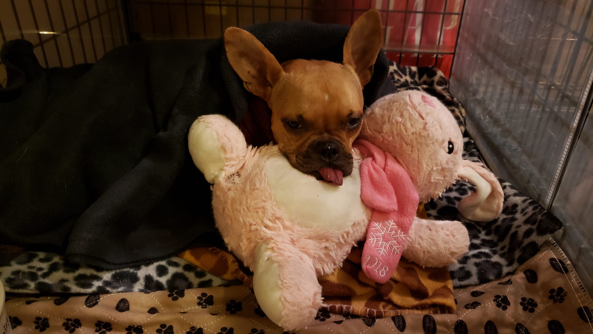
Her foster took exceptional care of her… She did not spoil her at all as you can see in this picture…
Only two days after surgery, her foster commented: “She is eating watered-down canned food, and lots of it. She is also drinking better. Hardly any sneezes. She doesn’t seem painful. I am amazed at how well she is doing honestly!! She seems surprised she can breathe through her nose and not sneezing.”
Four days after surgery, she kept improving: “Squirrel is now eating soft can food. No water added. She ate half a can this morning.”
Three weeks after surgery, we examined her mouth under sedation to assess the surgery site.
Thankfully, 100% of the surgery area “took.” The left side of the mouth (the pink area) still needed a bit more time to heal as nicely as the right, but I was reassured that everything worked out after the first surgery.
Squirrel’s foster concludes: “I cannot thank you enough for giving her this gift! She now has a new lease on life.”
Phil Zeltzman, DVM, DACVS, CVJ, FFcert.
Dr. Phil Zeltzman is a traveling veterinary surgeon in Pennsylvania & New Jersey. An award-winning author, he loves to share his adventures in practice along with information about vet medicine and surgery that can really help your pets. Dr. Zeltzman specializes in orthopedic, neurologic, cancer, and soft tissue surgeries for dogs, cats, and small exotics. By working with local family vets, he offers the best surgical care, safest anesthesia, and utmost pain management to all his patients. Sign up to get an email when he updates his blog, and follow him on Facebook, too!
Look at your dog’s head and predict health problems
The pet insurance branch of Nationwide recently released a report that shows that brachycephalic dogs have a higher risk for several diseases.
Brachycephalic dogs have a flat face. They tend to have a shorter skull, a shorter snout or muzzle and bulging eyes. Because of the shape of their skull, they often have breathing difficulties.
In an English Bulldog, the poster child for brachycephalic dogs, the nostrils are routinely very small (“stenotic nares”). This limits the flow of oxygen. In addition, a Bulldog can have a soft palate that is too long (the back part of the roof of the mouth), everted laryngeal saccules (small fleshy parts inside the larynx or voice box), and a small wind pipe (trachea). This blocks the flow of oxygen even more, which is why these dogs pant all the time. They are literally suffocating.
The smooshed face also leads to extra skin folds that can become irritated or infected.
The report shows a higher risk for skin diseases and eye problems.
Besides Bulldogs, brachycephalic dog breeds include the Affenpinscher, Boston terrier, boxer, Brussels griffon, old English bulldog, shih tzu, cavalier King Charles spaniel, dogue de Bordeaux, French bulldog, Japanese chin, Lhasa apso, mastiff, Brazilian mastiff, bull mastiff, English mastiff, Neapolitan mastiff, Pyrenean mastiff, Tibetan mastiff, Spanish mastiff, pekinese and pug.
What should you do?
All of these dogs can be wonderful pets, you just need to be aware of the health risks before you adopt them.
So what should you do if your dog has similar problems? The secret to avoid breathing problems later in life is to widen the nostrils as early as possible. Several articles* recommend at 3 to 4 months of age. The challenge is to operate on these dogs before the usual age to spay or neuter a dog. I agree 100% with these articles, early intervention is key.
As to the other issues, mainly skin diseases and eye problems, they are a whole different story. See your vet early on, before the problems go out of control.
Phil Zeltzman, DVM, DACVS, CVJ, Fear Free Certified
* Source: Here is a quote for an article about these patients: “It is recommended to perform rhinoplasty in puppies with this abnormality at 3 to 4 months of age to avoid progression to secondary changes, such as laryngeal collapse or pharyngeal edema.”
Dr. Phil Zeltzman is a traveling veterinary surgeon in Pennsylvania & New Jersey. An award-winning author, he loves to share his adventures in practice along with information about vet medicine and surgery that can really help your pets. Dr. Zeltzman specializes in orthopedic, neurologic, cancer, and soft tissue surgeries for dogs, cats, and small exotics. By working with local family vets, he offers the best surgical care, safest anesthesia, and utmost pain management to all his patients. Sign up to get an email when he updates his blog, and follow him on Facebook, too!