
Dr. Phil Zeltzman’s Blog
Fix your dog’s laryngeal paralysis with tie back surgery
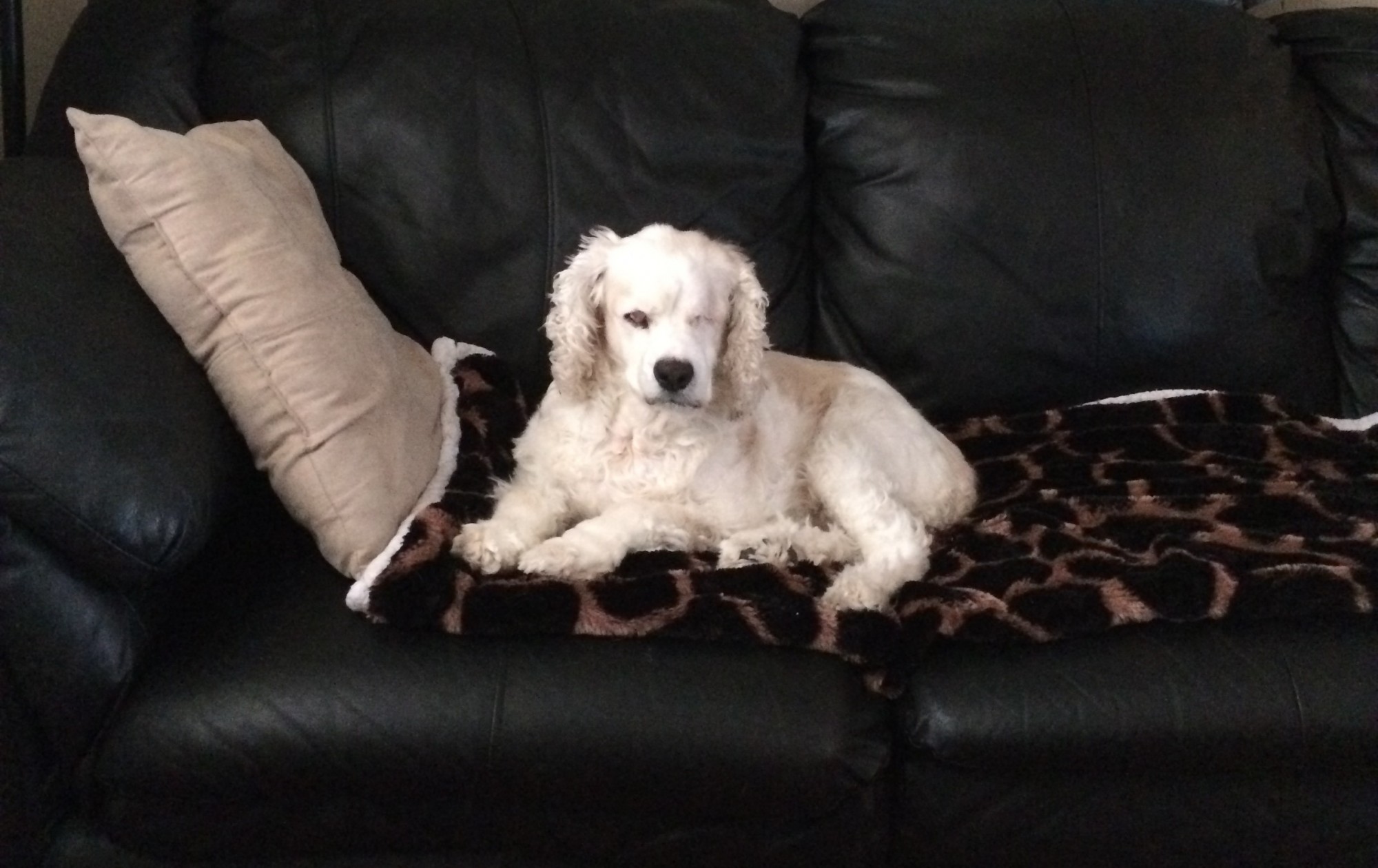 Matches is a 13 year old female Cocker Spaniel who, as you can see, rules the home from her royal couch.
She previously had to have her left eye removed because of glaucoma, but she was referred to me because she had difficulty breathing. Her family vet diagnosed her with an unusual condition in a Cocker: laryngeal paralysis, or lar par.
This was very surprising. It’s a common condition in Labradors, but rare in other breeds such as Cockers.
Lar par is a stressful ailment where the two folds of the larynx (or voice box) do not open and close as the patient breathes in and out. The folds remain closed – paralyzed – and the patient literally suffocates. This can be fixed with “tie back” surgery, which involves placing 2 strands of heavy nylon to open the left side of the larynx.
It’s a delicate surgery, but typically successful as it opens the airway so that the patient can breathe. Matches recovered very nicely and quickly from surgery at Blairstown Animal Hospital in New Jersey. So far, she is doing great.
Below, you can see a preop and postop video of the larynx.
In the “before” section, don’t be fooled by the movement at the bottom of the folds! These are the vocal cords, which do nothing for breathing. Look at the top of the folds, and you will see that they do not move, even when she tries to breathe in.
In the “after” portion, the left side of the throat (which appears on the right side of the screen!) is open to allow air from going in.
It’s very important for veterinary professionals to never assume! I was surprised to hear from my colleague that he had a Cocker with laryngeal paralysis, but sure enough, that’s what she had.
Dr. Phil Zeltzman is a traveling veterinary surgeon in Pennsylvania & New Jersey. An award-winning author, he loves to share his adventures in practice along with information about vet medicine and surgery that can really help your pets. Dr. Zeltzman specializes in orthopedic, neurologic, cancer, and soft tissue surgeries for dogs, cats, and small exotics. By working with local family vets, he offers the best surgical care, safest anesthesia, and utmost pain management to all his patients. Sign up to get an email when he updates his blog, and follow him on Facebook, too!
Never neglect a pet’s open wound
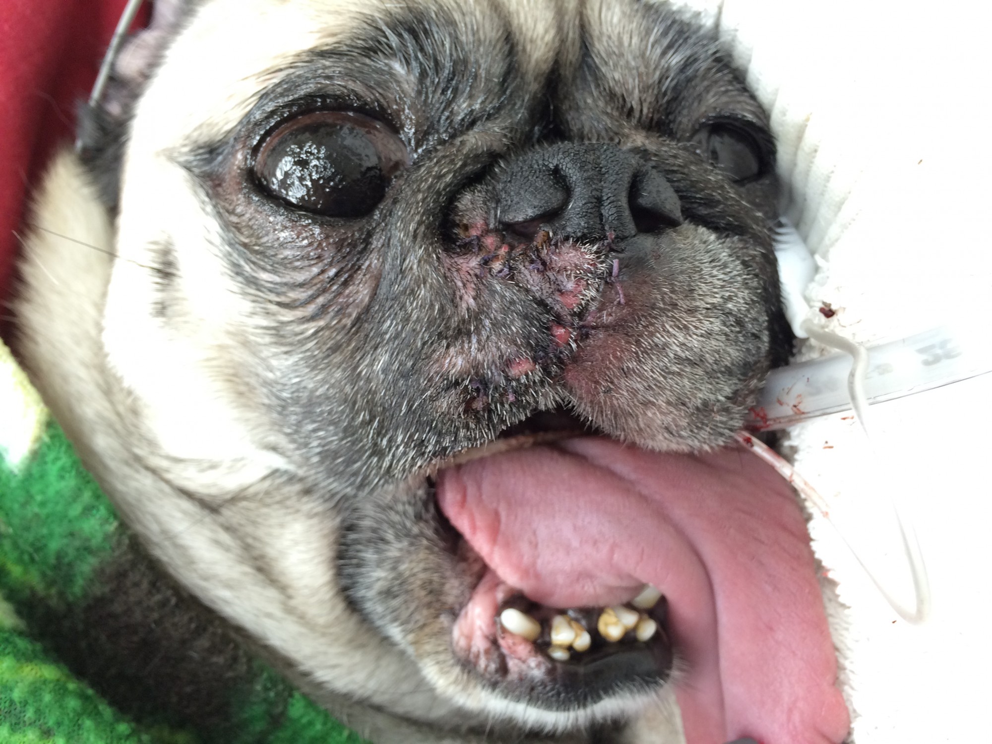
Quinton after surgery.
Quinton is an 8 year old Pug, who had a small open wound that would not heal despite antibiotics.
Quinton’s vet, instead of ignoring the wound, decided to test it. Under the microscope, the cells looked like Quinton could have a mast cell tumor! Mast cells are normal white blood cells that can occasionally cause a tumor. In fact, it’s one of the most common skin tumors. However, they typically appear as a lump or bump, not as an open wound.
I performed surgery at Barton Heights Veterinary Hospital and removed a very large portion of his right upper lip and small section of the left upper lip.
It is very important to remove enough tissue around a tumor in the hopes of getting it all.
The lab confirmed a mast cell tumor. There are 3 grades describing severity of the tumors: 1 is good, 3 is bad. Quinton had a grade 2 mast cell tumor, which we fortunately removed entirely.
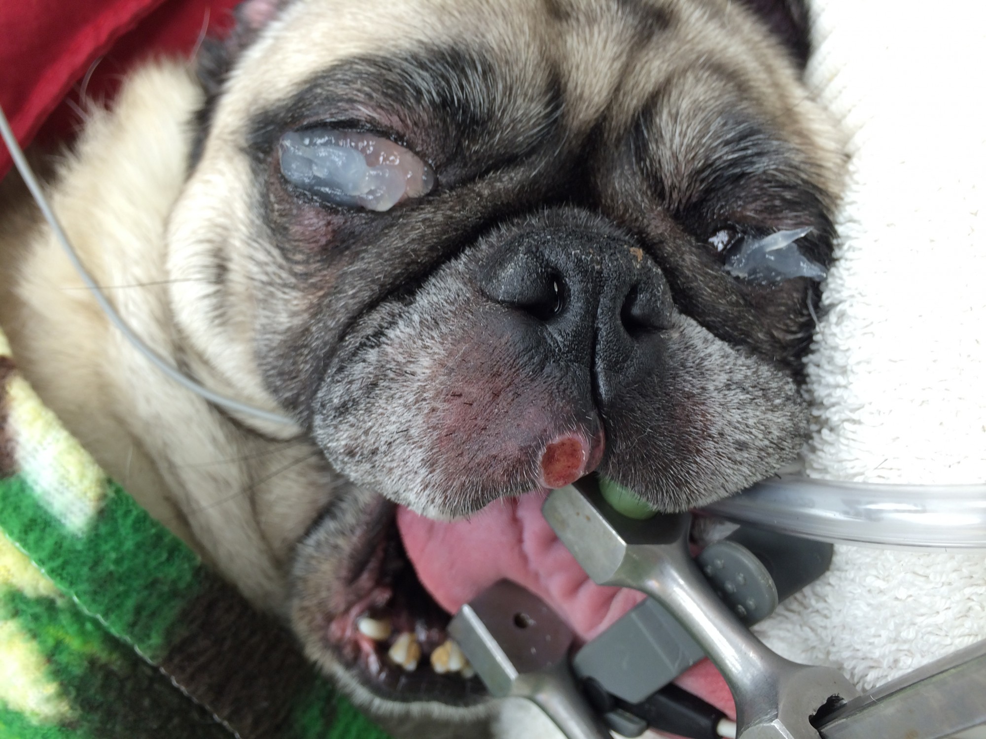
You can see the wound on Quinton’s lip.
Never neglect open wounds! You never know what they can be hiding.
Dr. Phil Zeltzman is a traveling veterinary surgeon in Pennsylvania & New Jersey. An award-winning author, he loves to share his adventures in practice along with information about vet medicine and surgery that can really help your pets. Dr. Zeltzman specializes in orthopedic, neurologic, cancer, and soft tissue surgeries for dogs, cats, and small exotics. By working with local family vets, he offers the best surgical care, safest anesthesia, and utmost pain management to all his patients. Sign up to get an email when he updates his blog, and follow him on Facebook, too!
Even ‘safe’ pet toys can be dangerous
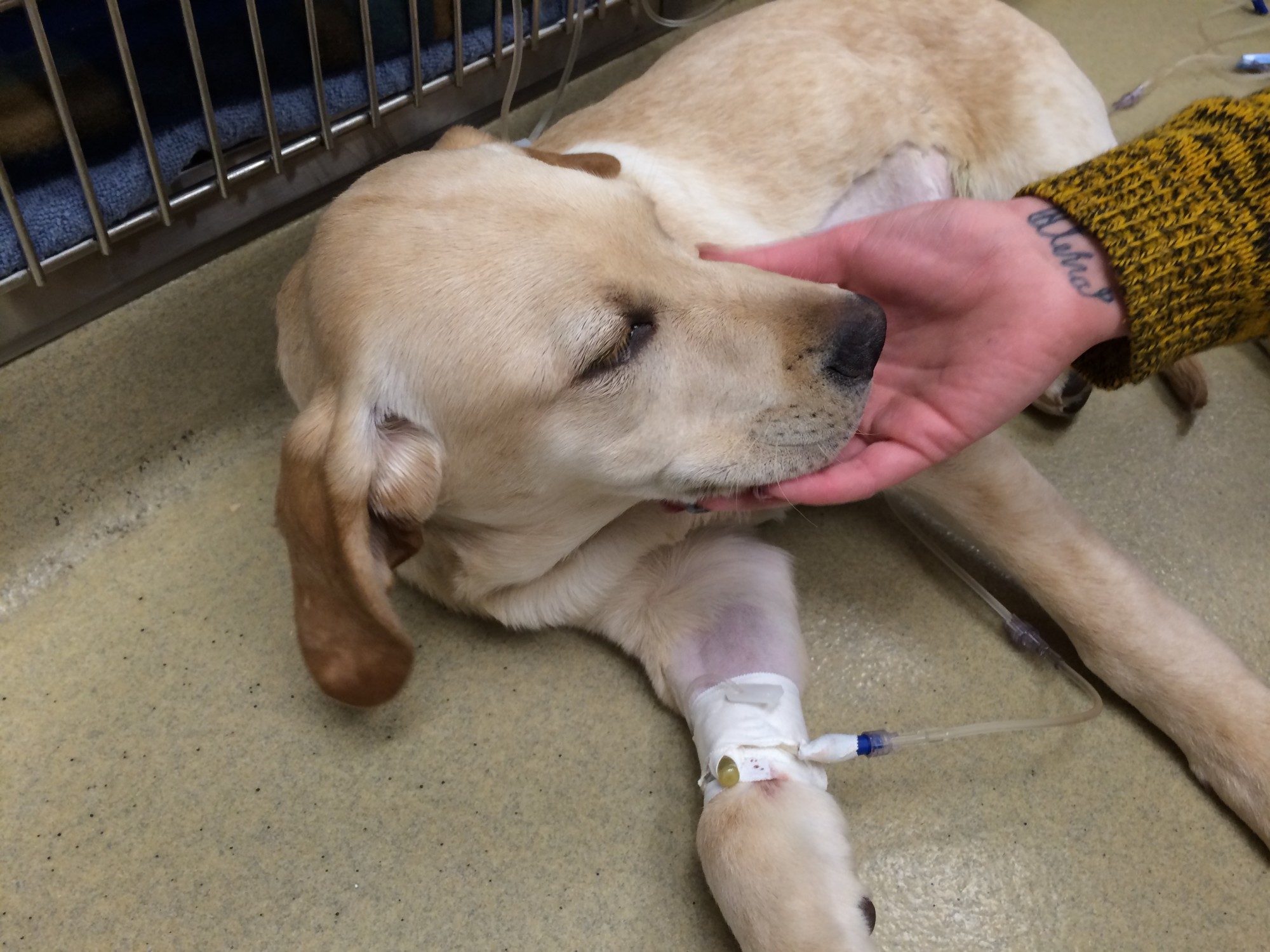 Trooper is a four month old Labrador puppy, who wasn’t feeling well. He was vomiting and not eating well.
His owners took him to an emergency clinic and x-rays revealed something that looked suspiciously like a foreign body. The veterinarian recommended surgery, which I performed at Berks Animal Emergency & Referral Center.
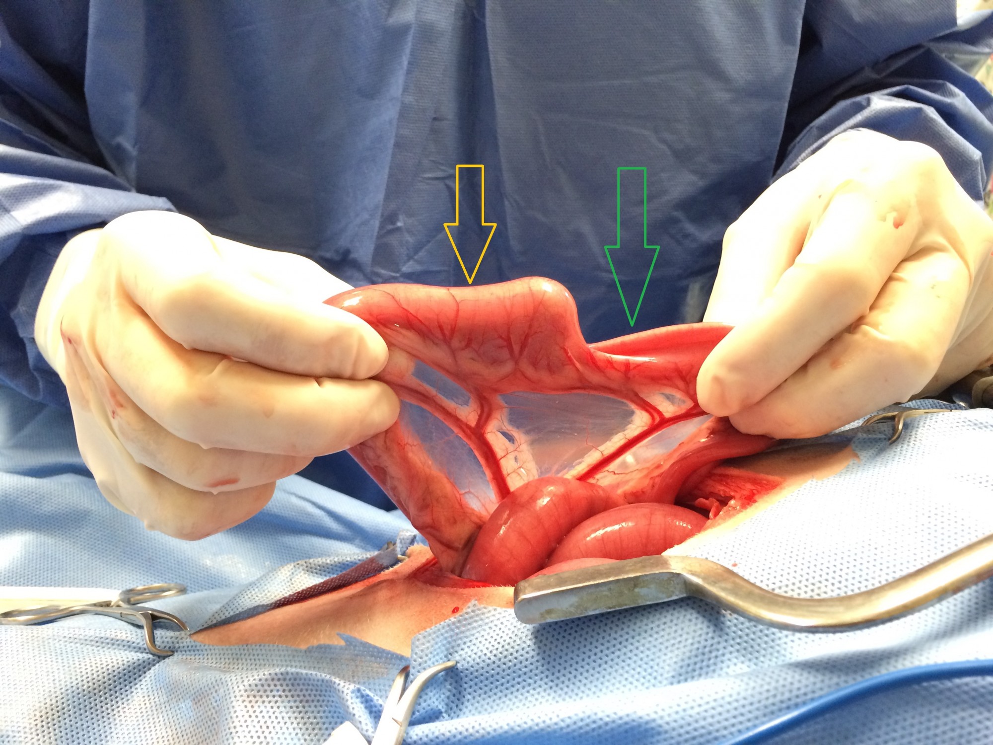
During surgery, I found hard foreign body stuck in the small intestine. The green arrow shows what the intestine should look like – small. The yellow arrow shows how the intestine is distended.
 The moral of the story? You should always monitor your pets’ toys! No toy is completely safe. Trooper’s owners did an excellent job of puppy-proofing the house: trash was secured, dirty laundry was inaccessible, and there was no way Trooper could get into something that would hurt him.
But they did not realize that a toy this hard could be chewed to pieces and swallowed. Lab puppies are masters at finding and eating things they shouldn’t!
Luckily, Trooper had a happy ending. He pulled through very well after anesthesia and surgery. In fact, he started eating just a few hours after waking up.
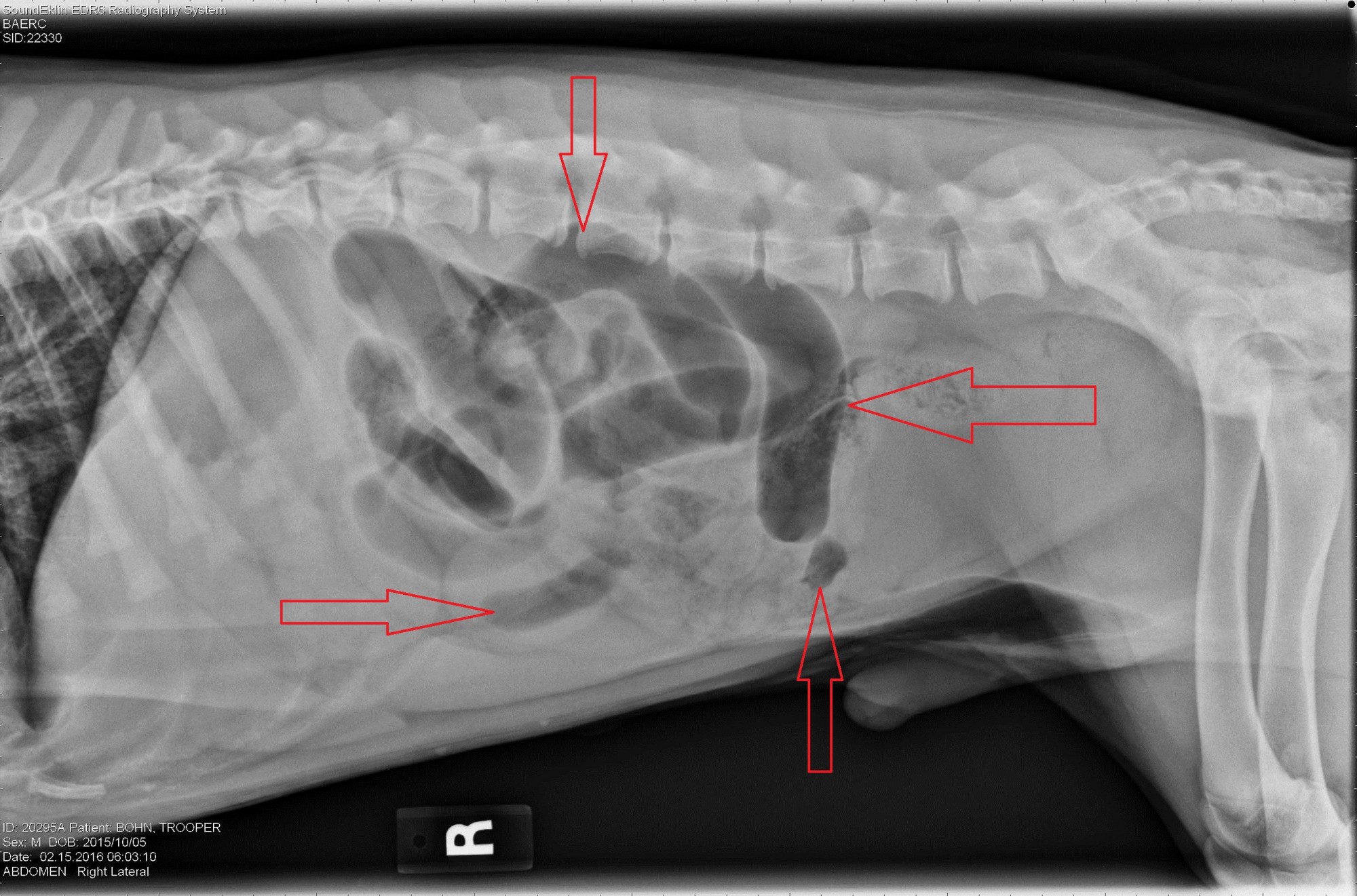 The x-ray shows typical gas bubbles of various sizes and shapes and a potential foreign body. |
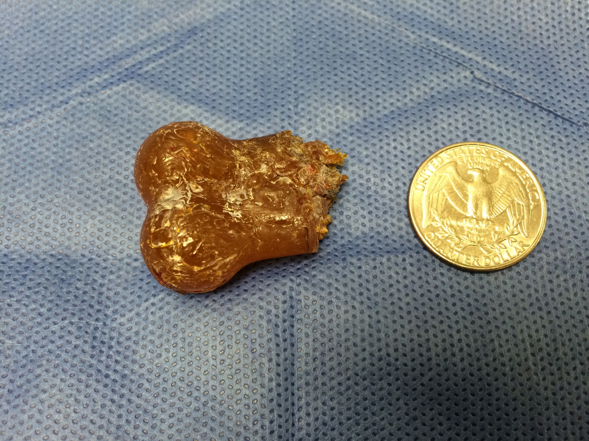 The toy that Trooper chewed up and swallowed. |
Dr. Phil Zeltzman is a traveling veterinary surgeon in Pennsylvania & New Jersey. An award-winning author, he loves to share his adventures in practice along with information about vet medicine and surgery that can really help your pets. Dr. Zeltzman specializes in orthopedic, neurologic, cancer, and soft tissue surgeries for dogs, cats, and small exotics. By working with local family vets, he offers the best surgical care, safest anesthesia, and utmost pain management to all his patients. Sign up to get an email when he updates his blog, and follow him on Facebook, too!
One incredibly lucky dog
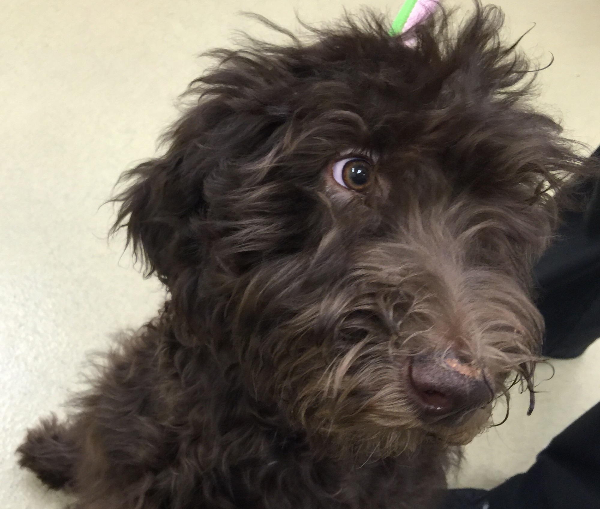 Marlee is an adorable four month old female Labradoodle, who was constantly licking her lips.
Her family took her to the emergency vet whose first thought was something might be stuck in Marlee’s mouth or throat. An exam under sedation confirmed the suspicion: there was a string wrapped around her tongue. He gently pulled on it, but it was stuck.
Marlee had a classic “string foreign body” or “linear foreign body.” It typically goes down into the stomach or intestine. Once it reaches the intestine, the string can cut into it and cause leakage of its content. This causes peritonitis, which can be deadly.
I performed Marlee’s surgery at Berks Animal Emergency and Referral Center. Marlee’s belly was opened up in surgery. Her stomach was full of food. Luckily, the intestine did not seem to be affected, which meant the string was stuck somewhere in the stomach.
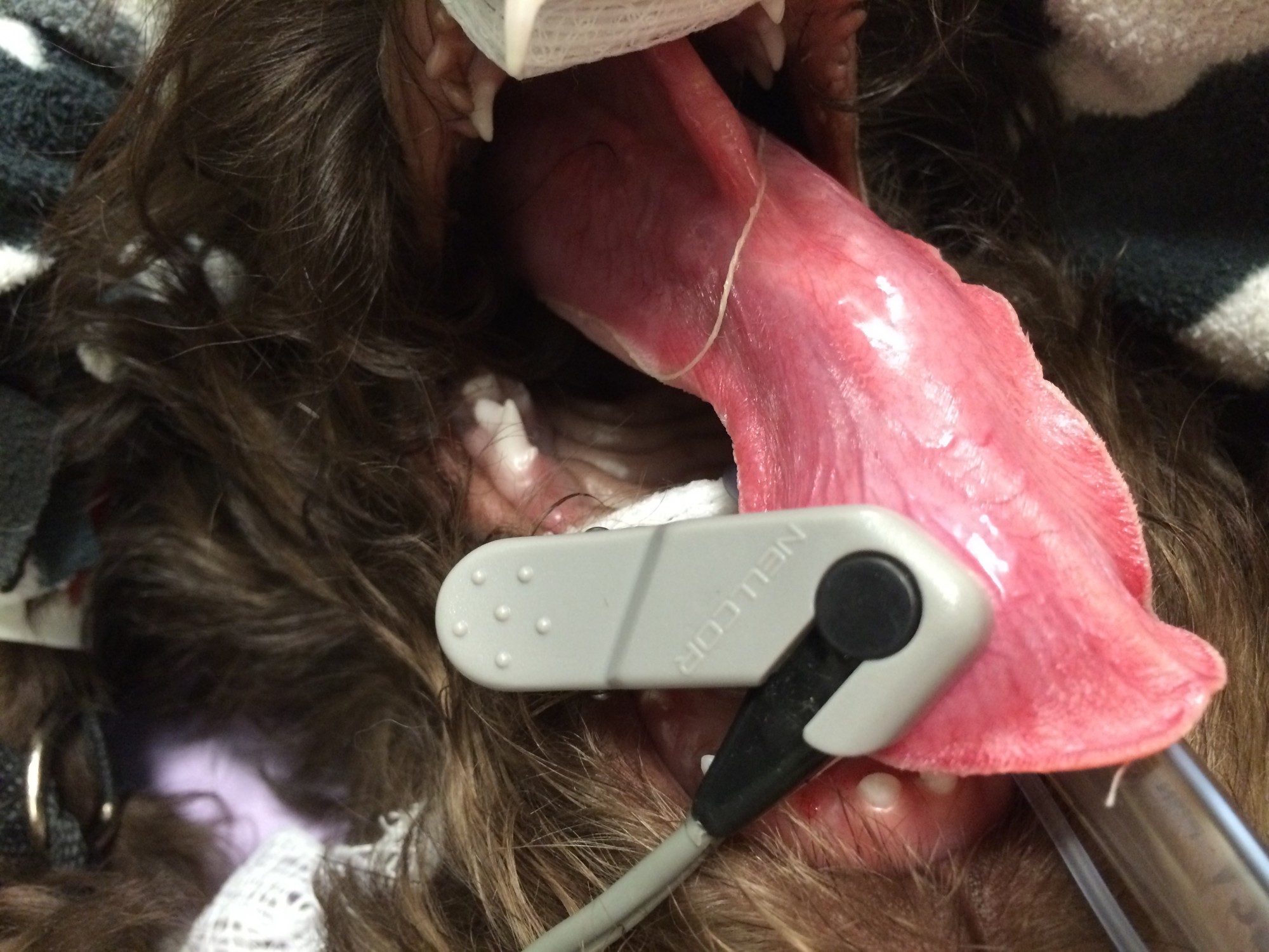
You can see the string wrapped around Marlee’s tongue before it disappears down her esophagus and into the stomach.
I felt around, but it was impossible to recognize anything because of the amount of food. I asked the anesthesia nurse to gently pull on the string from inside the mouth. It was stuck. If the nurse couldn’t pull the string out, our only other option was to open the stomach, and dig around until the string would be found.
At the last second, just before I cut into the stomach, something slipped between my fingers and I distinctly felt a round foreign body slide from the stomach into the esophagus (the tube between the mouth and the stomach).
This stroke of luck allowed the anesthesia nurse to gently pull on the string. Little by little, she managed to pull two feet worth of string through the mouth!
Marlee recovered beautifully! She woke up smoothly after anesthesia, and by the time of her staple remove two weeks post-op, she was completely back to normal.
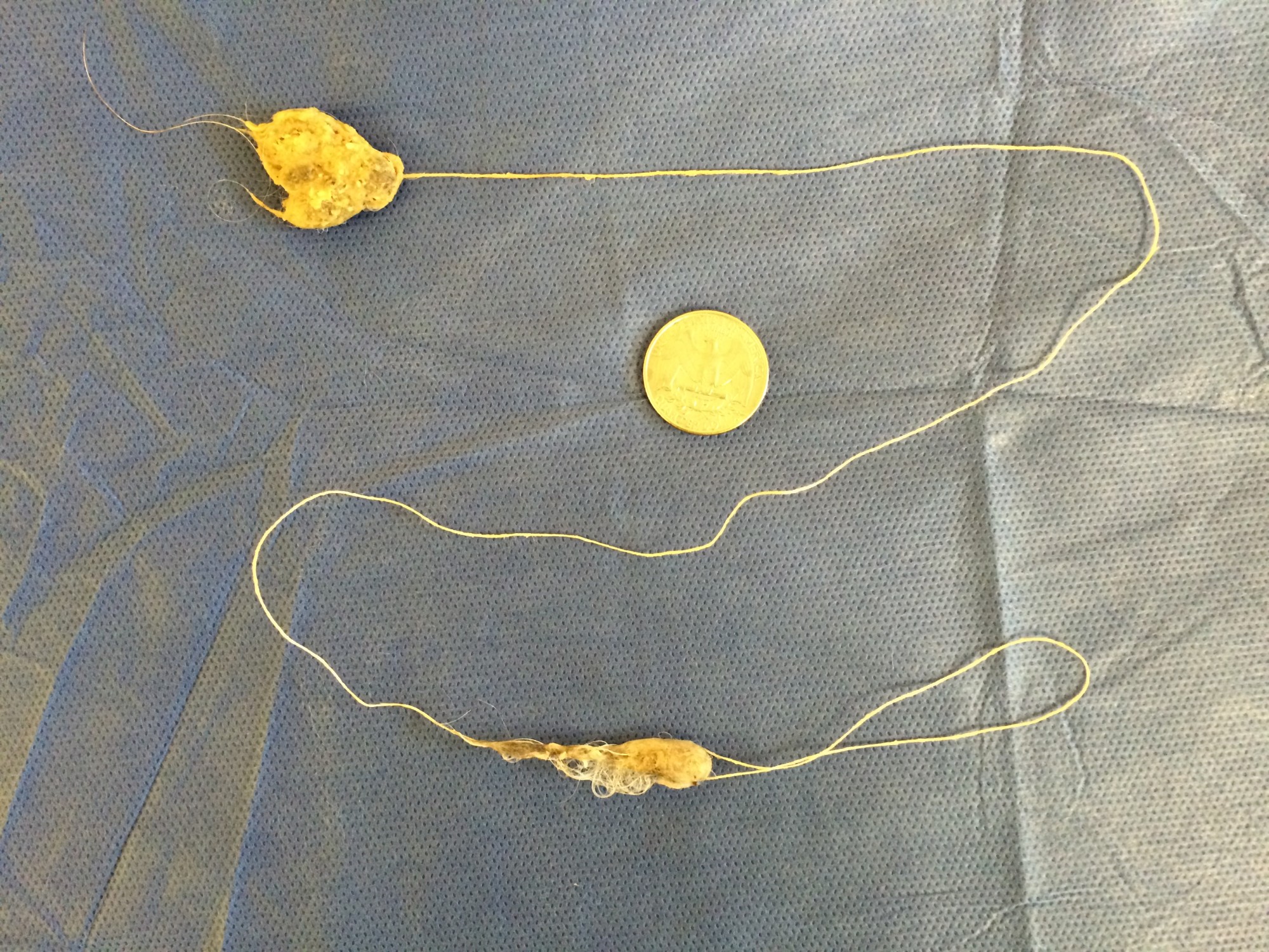
The offending string was two feet long.
Dr. Phil Zeltzman is a traveling veterinary surgeon in Pennsylvania & New Jersey. An award-winning author, he loves to share his adventures in practice along with information about vet medicine and surgery that can really help your pets. Dr. Zeltzman specializes in orthopedic, neurologic, cancer, and soft tissue surgeries for dogs, cats, and small exotics. By working with local family vets, he offers the best surgical care, safest anesthesia, and utmost pain management to all his patients. Sign up to get an email when he updates his blog, and follow him on Facebook, too!
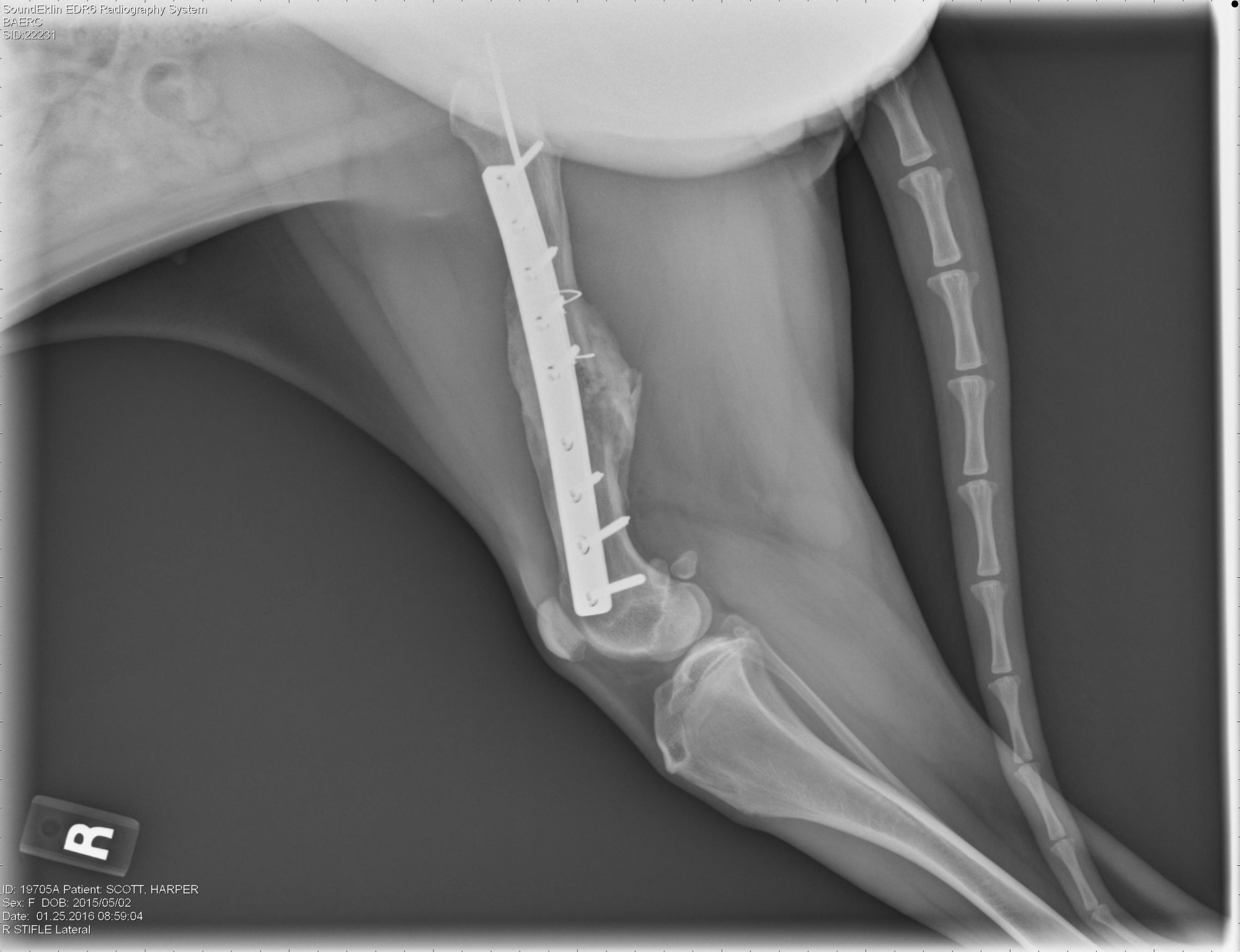
Shattered femur causes problems for German Shepherd
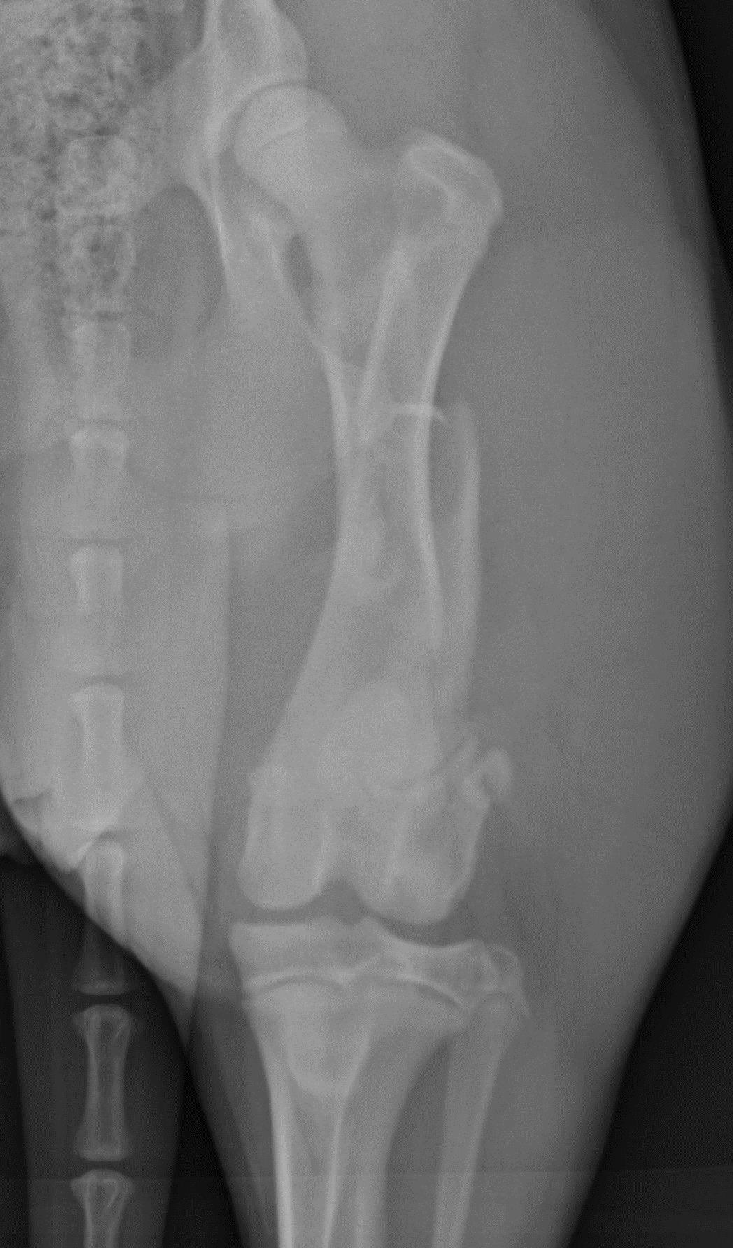
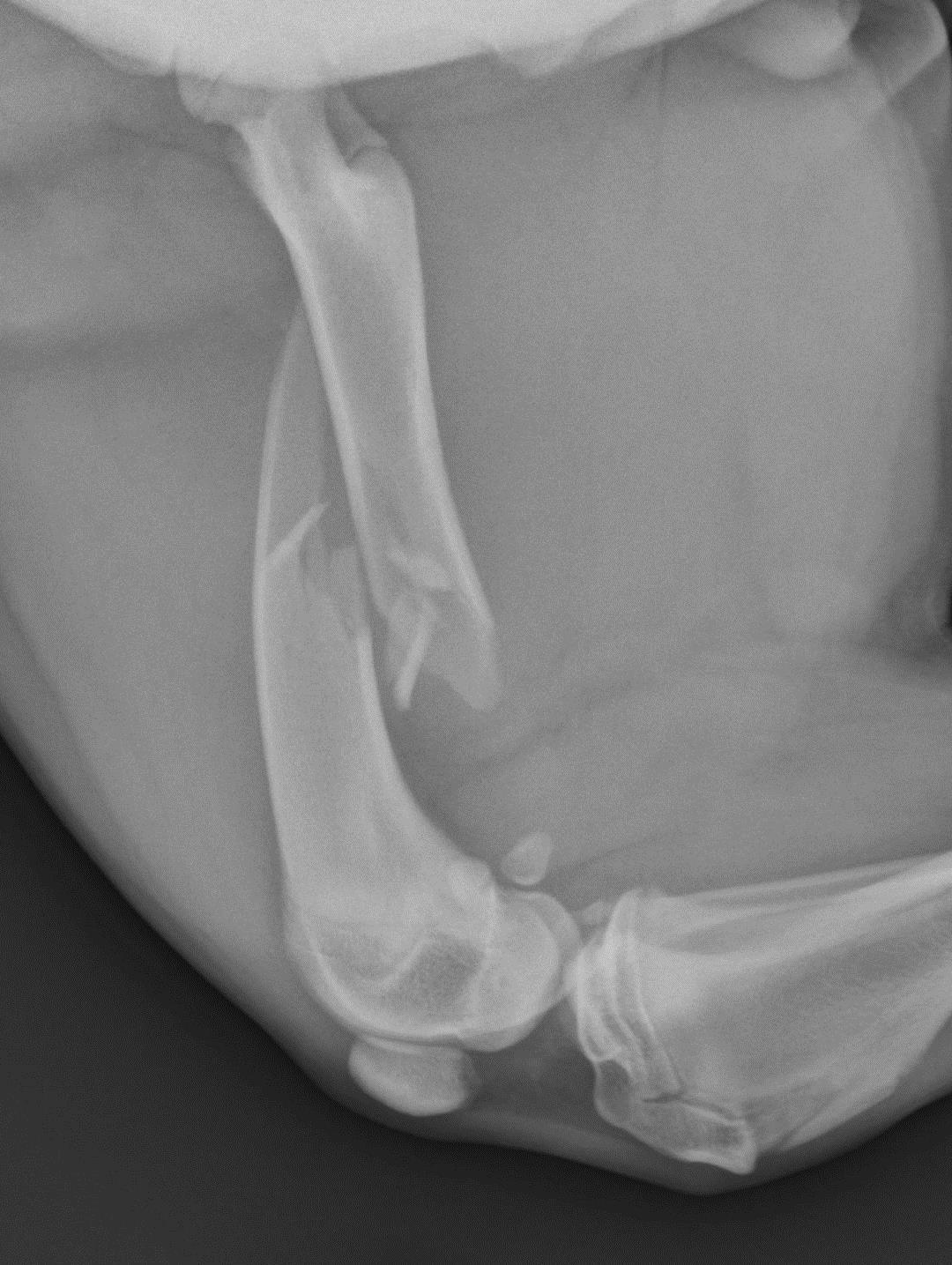
 Harper, a 6 month old female German Shepherd, had a small problem.
Lots of small problems actually.
Her femur, or thigh bone, was shattered. You can see two large pieces at the break, and multiple small pieces in the middle.
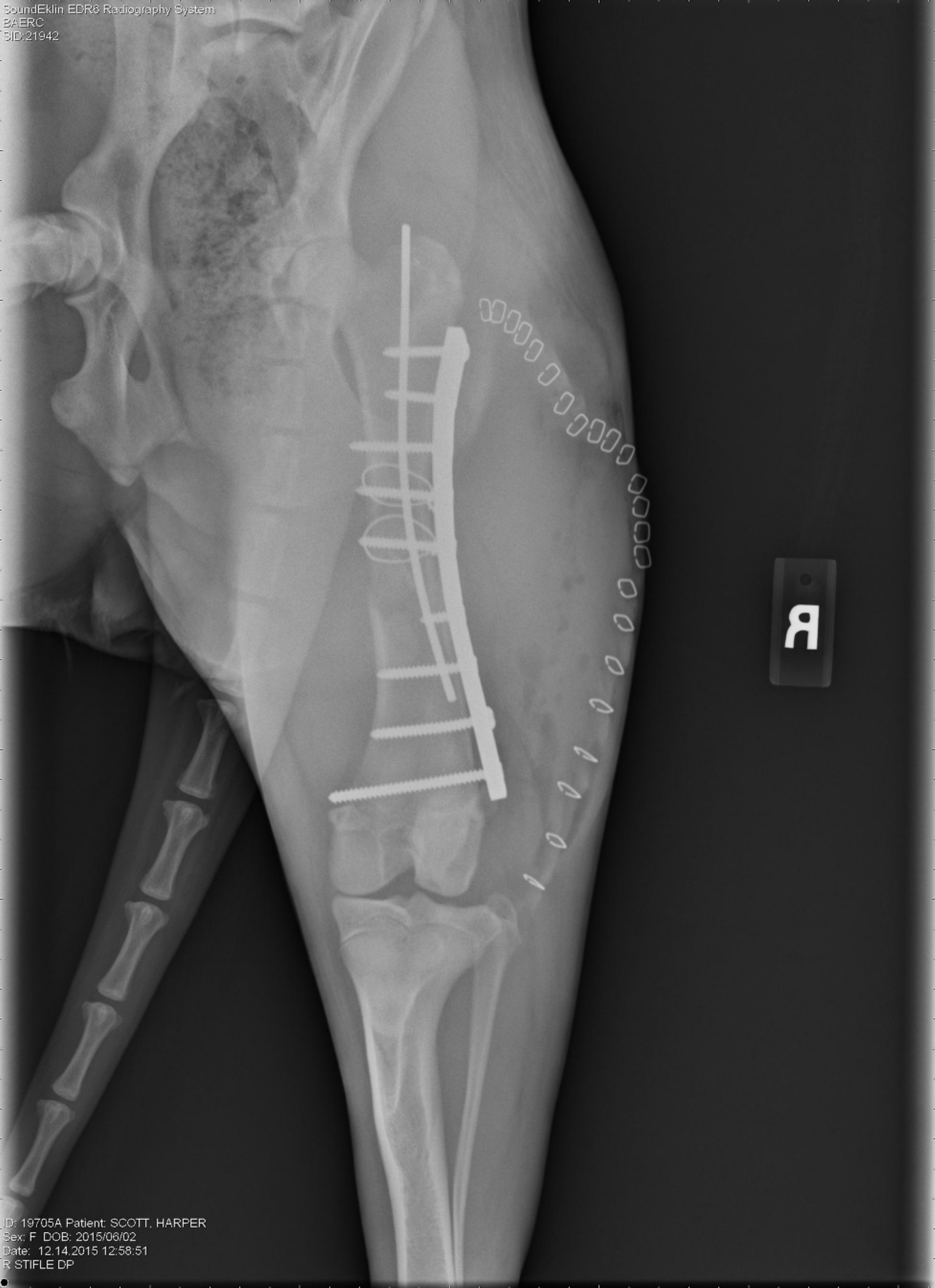
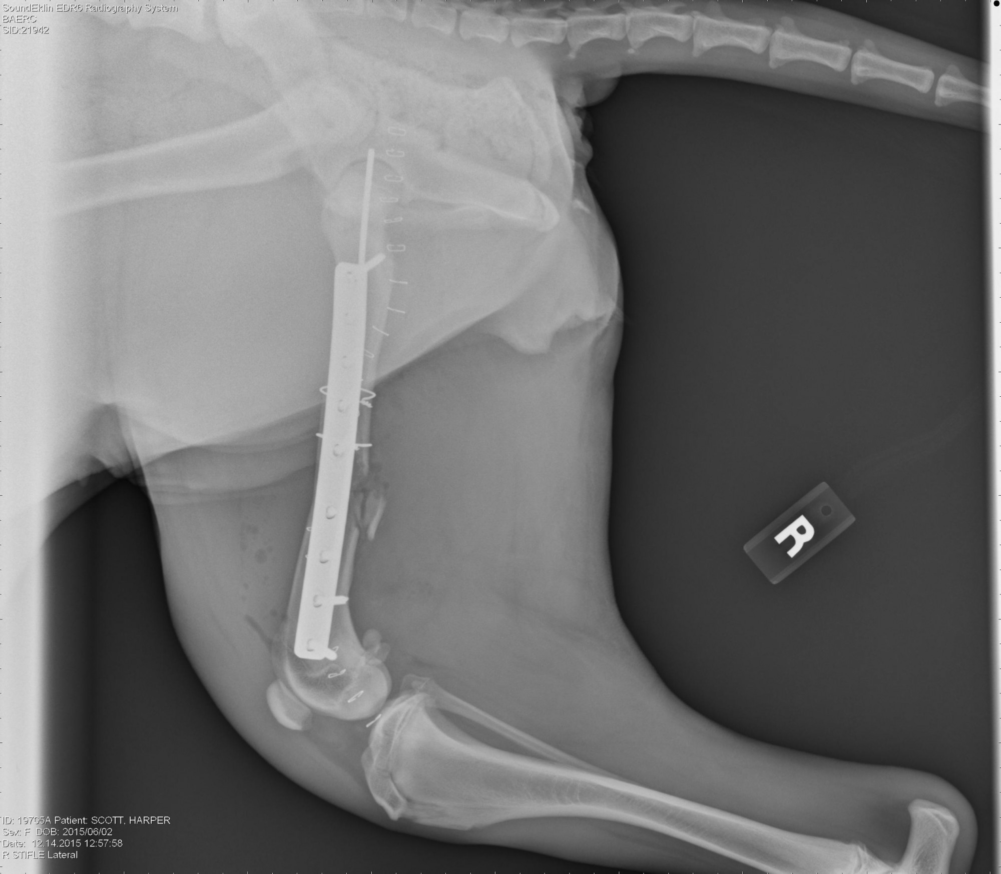
I performed a surgical repair at Berks Animal and Emergency Center & Referral Center, using 1 pin, 2 wires, 9 screws, a stainless steel plate, and a bone graft! You can see the fractures and the repairs in the x-ray images below.
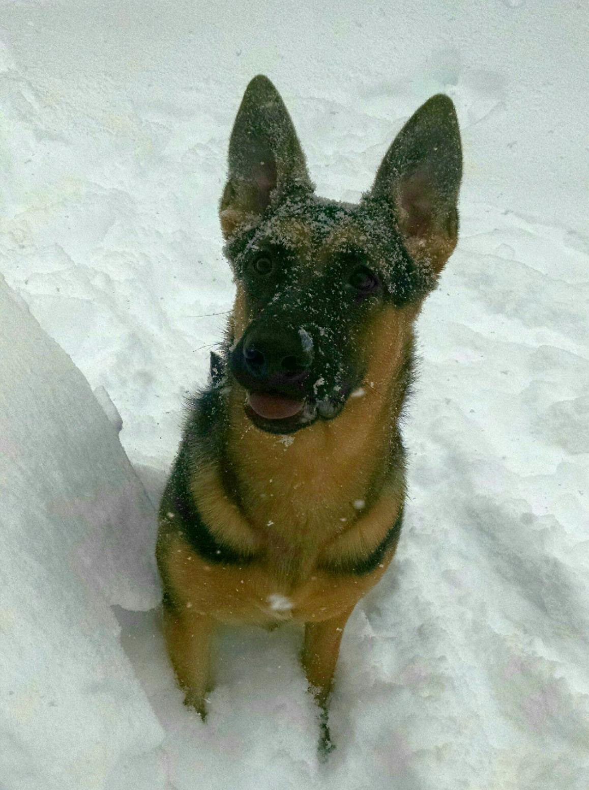
After 6 weeks of strict confinement, it was time to play in the snow!
 A view of the femur, immediately before surgery. |
 Another preop view of the femur. |
 Here is Harper immediately after surgery. |
 A lateral post-surgical view. |
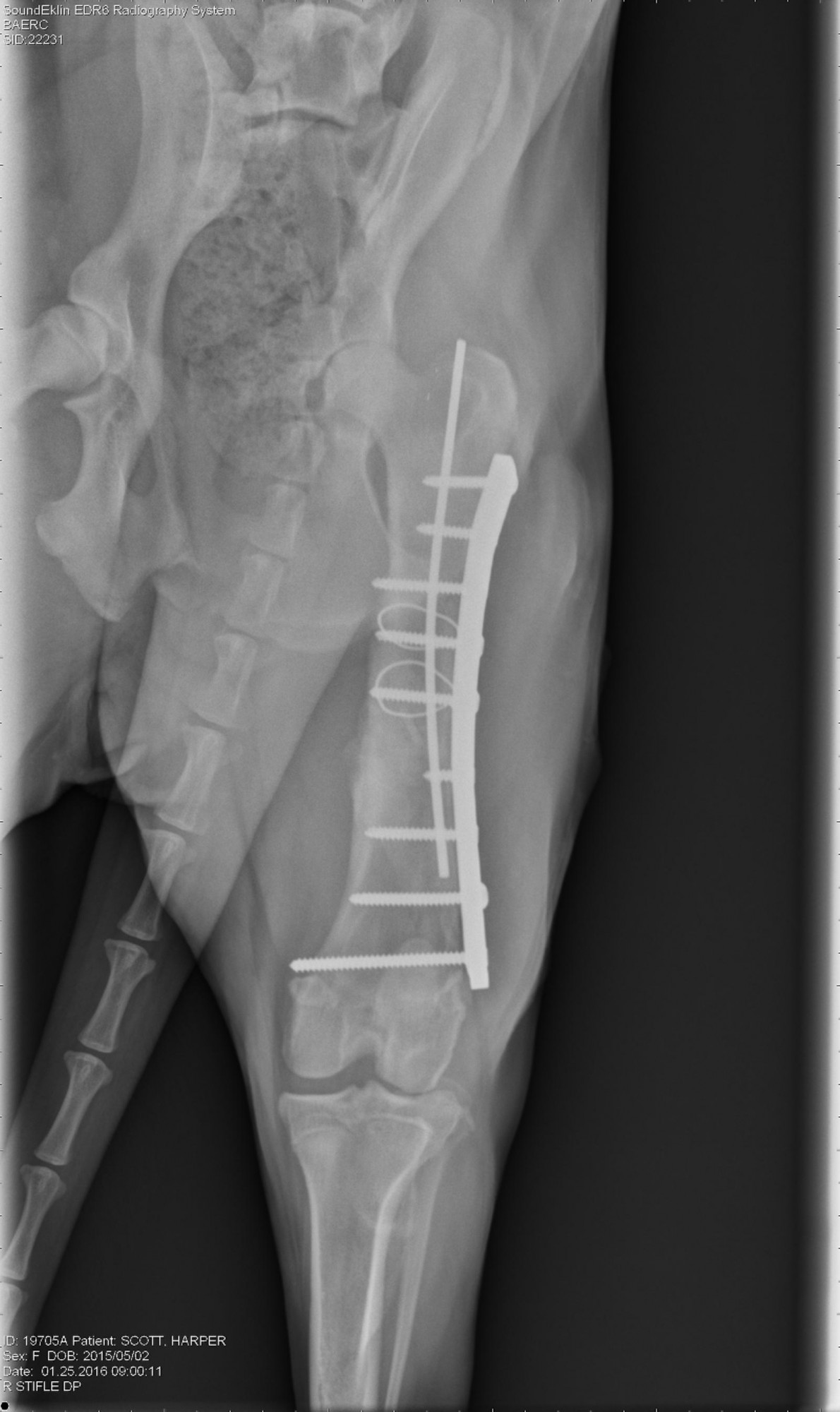 An x-ray six weeks after surgery. |
 Six weeks after surgery, Harper’s x-rays look much better! |
Dr. Phil Zeltzman is a traveling veterinary surgeon in Pennsylvania & New Jersey. An award-winning author, he loves to share his adventures in practice along with information about vet medicine and surgery that can really help your pets. Dr. Zeltzman specializes in orthopedic, neurologic, cancer, and soft tissue surgeries for dogs, cats, and small exotics. By working with local family vets, he offers the best surgical care, safest anesthesia, and utmost pain management to all his patients. Sign up to get an email when he updates his blog, and follow him on Facebook, too!