Dr. Phil Zeltzman’s Blog
Valentine, the rescued kitten with a broken leg
Valentine, a 7 month old kitten, was rescued after someone noticed that he (yes he’s a boy), that he was not putting any weight on his right back leg. X-rays showed that he had a fracture at the very end of the right thigh bone (see X-ray below).
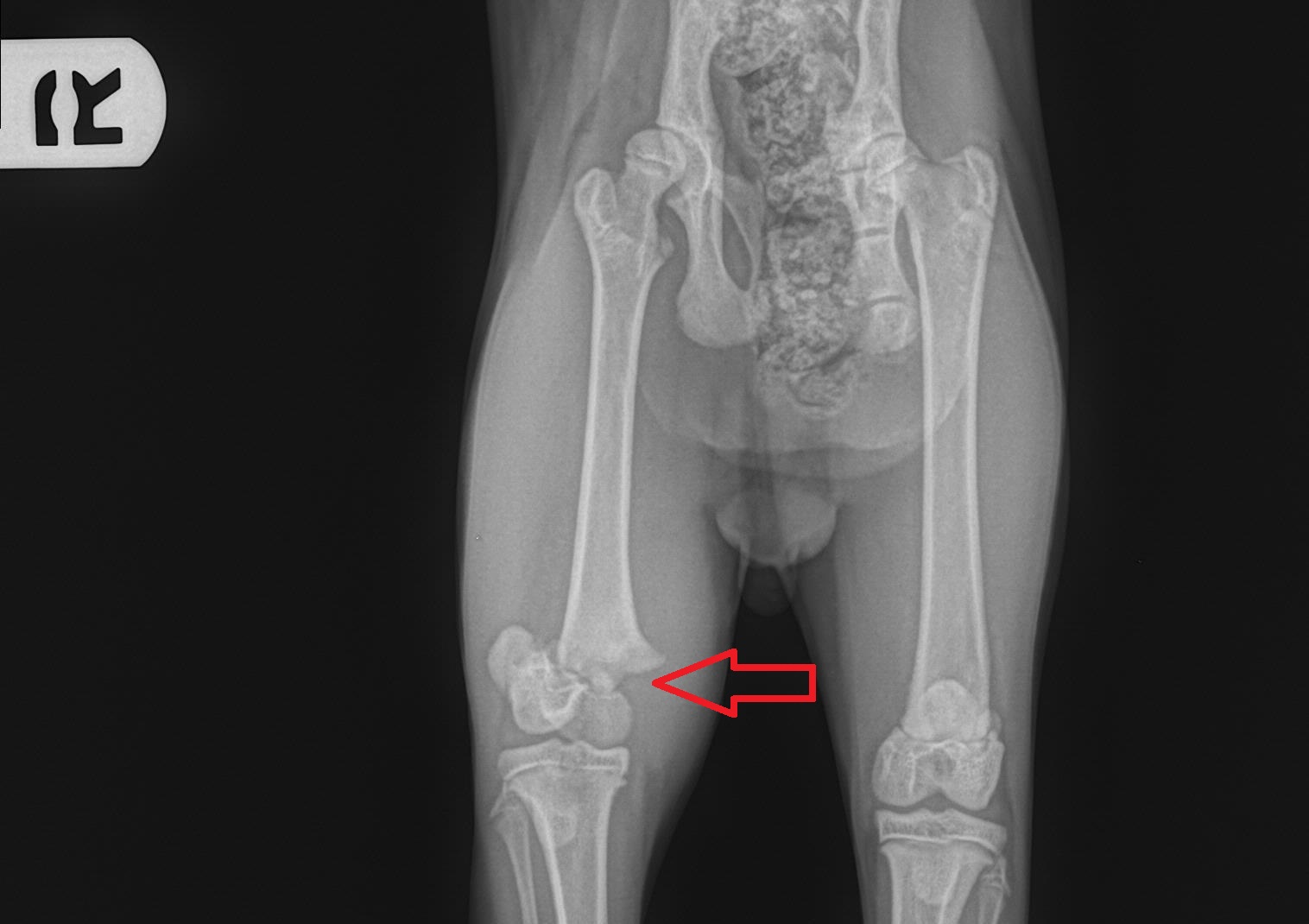
An exam showed that the bone had already healed. Yet the X-ray showed that the bone was grossly misaligned (see red arrow).
So we took Valentine to surgery with 2 options in mind:
. If the fracture could be repaired nicely, then we would fix the bone.
. If a good outcome could not be reached, we would sacrifice the leg (a nice way to say that we would amputate the leg).
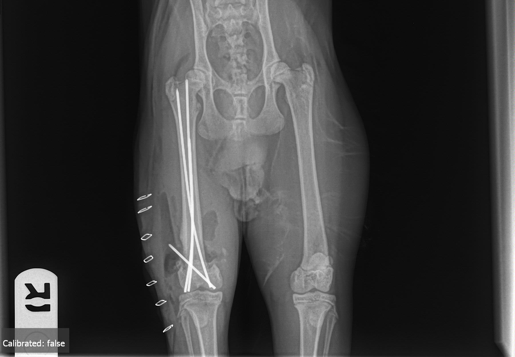
In surgery, I confirmed that the bone had healed in a crooked way. The kneecap was completely dislocated as a consequence. We had to “rebreak the bone”. I then removed a bunch of scar tissue, realigned the bone, and repaired it with 4 metal pins (see postop X-ray below).
Then I repaired the dislocated kneecap.
After 6 weeks of strict rest and physical therapy, Valentine should be able to find a furever home and enjoy a happy life.
Dr. Phil Zeltzman is a traveling veterinary surgeon in Pennsylvania & New Jersey. An award-winning author, he loves to share his adventures in practice along with information about vet medicine and surgery that can really help your pets. Dr. Zeltzman specializes in orthopedic, neurologic, cancer, and soft tissue surgeries for dogs, cats, and small exotics. By working with local family vets, he offers the best surgical care, safest anesthesia, and utmost pain management to all his patients. Sign up to get an email when he updates his blog, and follow him on Facebook, too!
The amazing story of Rori and her lung tumor
In June 2016, Rori, a 12 year old Westie, had visited the emergency hospital because she wasn’t feeling well. She was depressed and coughing.
Chest X-rays revealed a mass in her left lung. She did well for a few months, until her next set of X-rays in February 2017.
They revealed that the mass almost doubled in size (the round mass is at the end of the arrow on the X-ray below)!

Because of the high risk of lung cancer, surgery was recommended to remove the part of the lung that contained the tumor.
The odds were definitely against us: about 85% of lung tumors are cancerous. In addition, lung cancer is aggressive: the average survival after surgery alone is around 1 year. Lastly, a tumor that doubles in size in a few months is a definite concern.
Despite the odds, Rori’s owner wanted to provide the best possible quality of life for her dog and elected to move on with surgery.
To remove the tumor, we don’t “crack the ribs” like they say on TV. Rather, we go between 2 ribs. After entering the chest, the mass was found exactly where the X-rays showed. The mass was approximately the size of a nickel (the coin shown below is a quarter).
Below is a video during surgery. Warning !!!
THIS VIDEO IS VERY GRAPHIC, SO IT IS NOT FOR THE FAINT OF HEART !!!
That part of the lung (called a lung lobe) was sutured and removed. We did not see any other problem, so the chest was sutured closed.
The mass is visible on top of a lung lobe
The evening of the surgery, Rori ate her first meal !
The next day, she felt much better and was amazingly comfortable. She eventually went home.
A few days later, the biopsy report came back.
Despite the very high odds of cancer, unbelievably, the mass was… benign!
This was fantastic news. It meant that the mass should not affect Rori’s lifespan at all.
Few pet owners would have put their 12 year old dog through open chest surgery, knowing the risk of cancer was so high. But Rori’s owner chose surgery… and was rewarded in an unexpected way.
APRIL 2017 UPDATE:
It’s been 6 weeks since surgery, and Rori is doing very well during her recovery.
I explained to her owner how to slowly increase her activity level over the next month.
Both are thrilled with the idea!
Dr. Phil Zeltzman is a traveling veterinary surgeon in Pennsylvania & New Jersey. An award-winning author, he loves to share his adventures in practice along with information about vet medicine and surgery that can really help your pets. Dr. Zeltzman specializes in orthopedic, neurologic, cancer, and soft tissue surgeries for dogs, cats, and small exotics. By working with local family vets, he offers the best surgical care, safest anesthesia, and utmost pain management to all his patients. Sign up to get an email when he updates his blog, and follow him on Facebook, too!
An early spay could save your pet from emergency surgery
 Dakota, a 7-year-old female Boxer, was brought to her family vet because she hadn’t been acting herself for about two weeks. She was drinking more, eating less, and seemed weak.
X-rays revealed that Dakota had a very large uterus. Her classic symptoms and the X-rays confirmed that Dakota had a pyometra. This means that her uterus was filled with a large amount of pus.
This can be deadly.
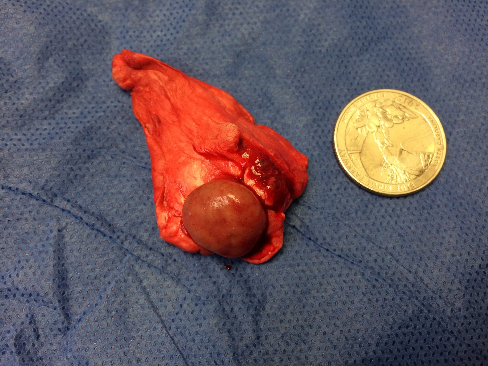
Emergency surgery was performed that same evening at Brodheadsville Veterinary Clinic. Removing a pyometra is essentially a complicated spay procedure. The main difference is that the uterus is huge and filled with infected fluid. Care must be taken to remove the infected uterus without any spillage to the rest of the belly. After surgery, the uterus weighed in at 7 pounds.
You can see a video of part of the surgery below. Just know that it contains graphic footage of a surgical procedure, so if you’re sensitive to that you may want to skip the video.
Dakota recovered quickly from surgery and anesthesia, and started eating and drinking normally shortly after that. She was back to her lovable self in one week.
Spaying your pets is essential! If you spay your cat or dog at an early age, you help reduce the risk of uterine cancer, ovarian cancer, and pyometra. Plus, spaying your pet before the first heat cycle virtually eliminates the risk of mammary tumors (including breast cancer).
If you are not planning to breed your female pet, please plan to have her spayed before she ends up in a life-threatening situation!
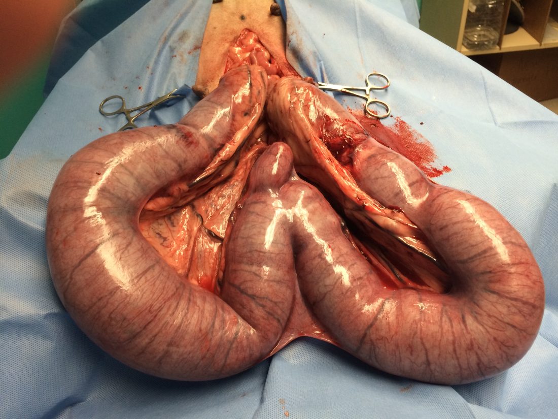 Dakota’s uterus during surgery |
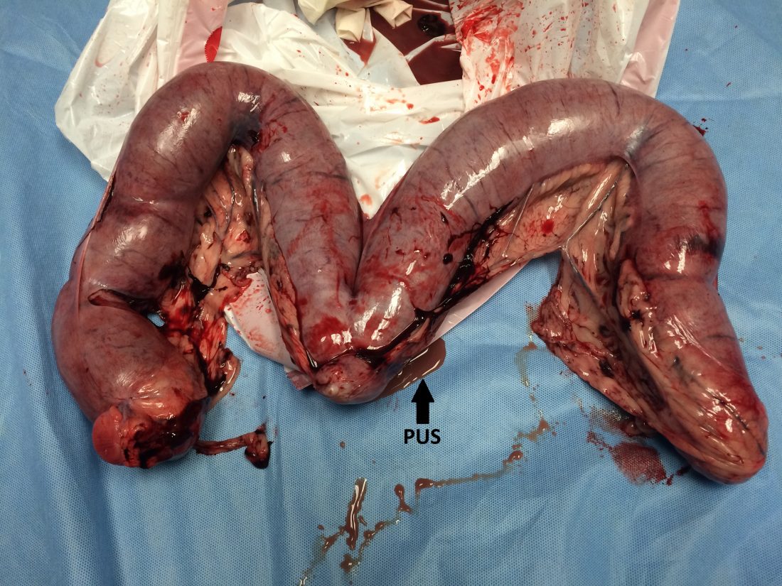 A post-op picture of the removed uterus. |
Dr. Phil Zeltzman is a traveling veterinary surgeon in Pennsylvania & New Jersey. An award-winning author, he loves to share his adventures in practice along with information about vet medicine and surgery that can really help your pets. Dr. Zeltzman specializes in orthopedic, neurologic, cancer, and soft tissue surgeries for dogs, cats, and small exotics. By working with local family vets, he offers the best surgical care, safest anesthesia, and utmost pain management to all his patients. Sign up to get an email when he updates his blog, and follow him on Facebook, too!
Chow needs surgery to fix mysterious swelling
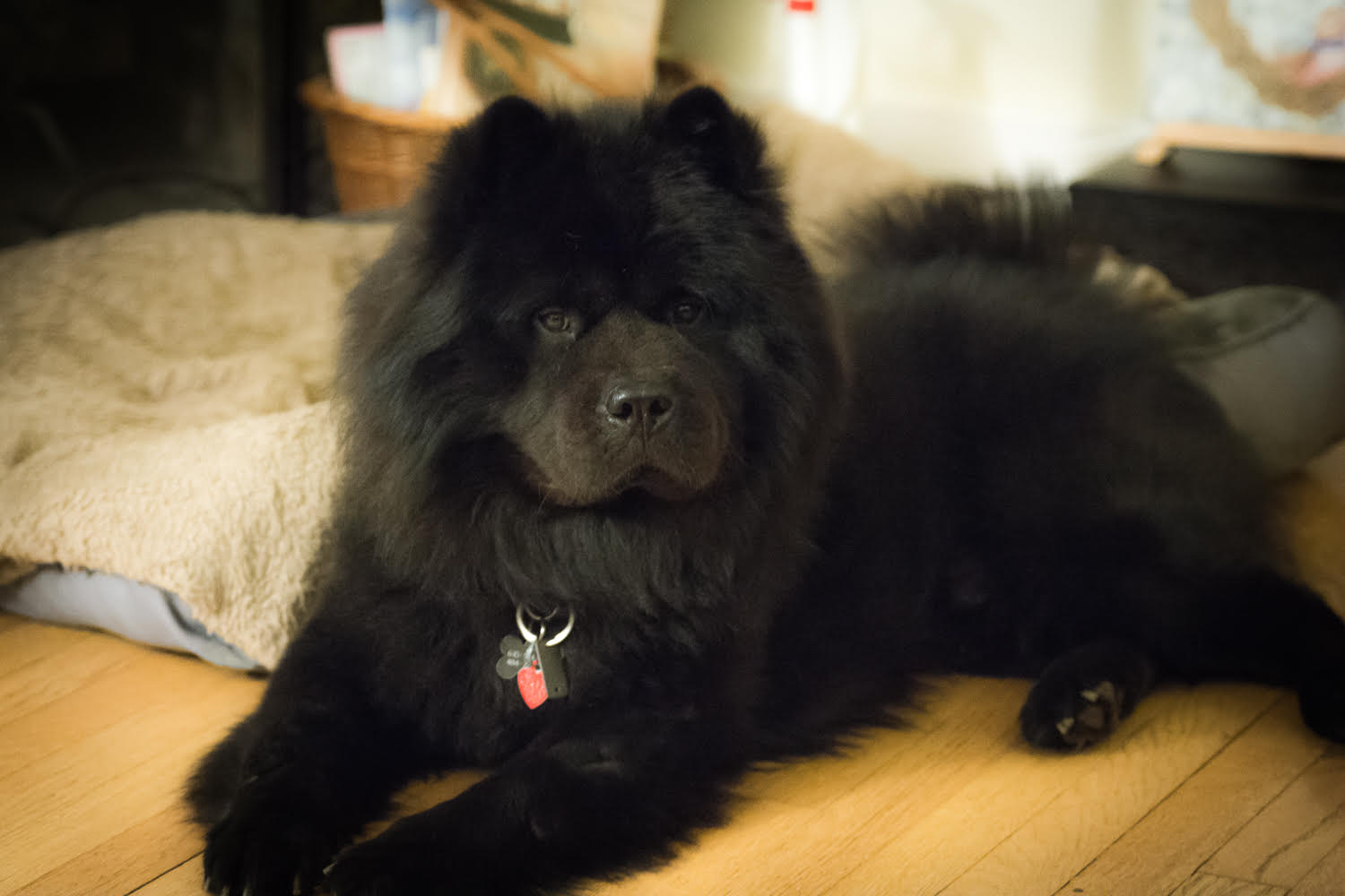
Photo courtesy of MHNG Photography.
Maxwell, a three-year-old male Chow, was relinquished from a home where he spent most of his time in a cage in a dark basement. After his rescue, he spent some time with a foster family, then was recently adopted by his forever family.
His new owner noticed a swelling on Maxwell’s cheek and took him to his family veterinarian. The vet was able to feel a firm swelling on the right side of the head, behind the angle of the jaw. In that area, any swelling should be suspicious for an enlarged lymph node. This can be a sign of a type of cancer called lymphoma.
The vet performed a needle aspirate, which thankfully confirmed a sialocele, a pocket of thick saliva. We typically never find out the reason for this condition. It’s assumed to be caused by trauma or blockage of the tiny canal that carries saliva from the salivary gland to the mouth.
The sialocele was carefully removed at HanoverView Animal Hospital. It’s a delicate surgery, because a sialocele is a very fragile structure that can tear easily. In addition, there are several important blood vessels and nerves in that area. Maxwell did very well under anesthesia and during recovery.
A week later, the biopsy confirmed a benign sialocele. Removing it will have no consequences for Maxwell because there are many salivary glands.
It is very important to pet your dog or cat thoroughly and regularly so you can notice any new lump or bump. Maxwell is a furry dog with a thick coat, and he is lucky his owner felt the swelling.
Any swelling or lump should be investigated by your family vet as soon as you notice it. Three important questions should be answered: should the mass be tested, removed and biopsied?
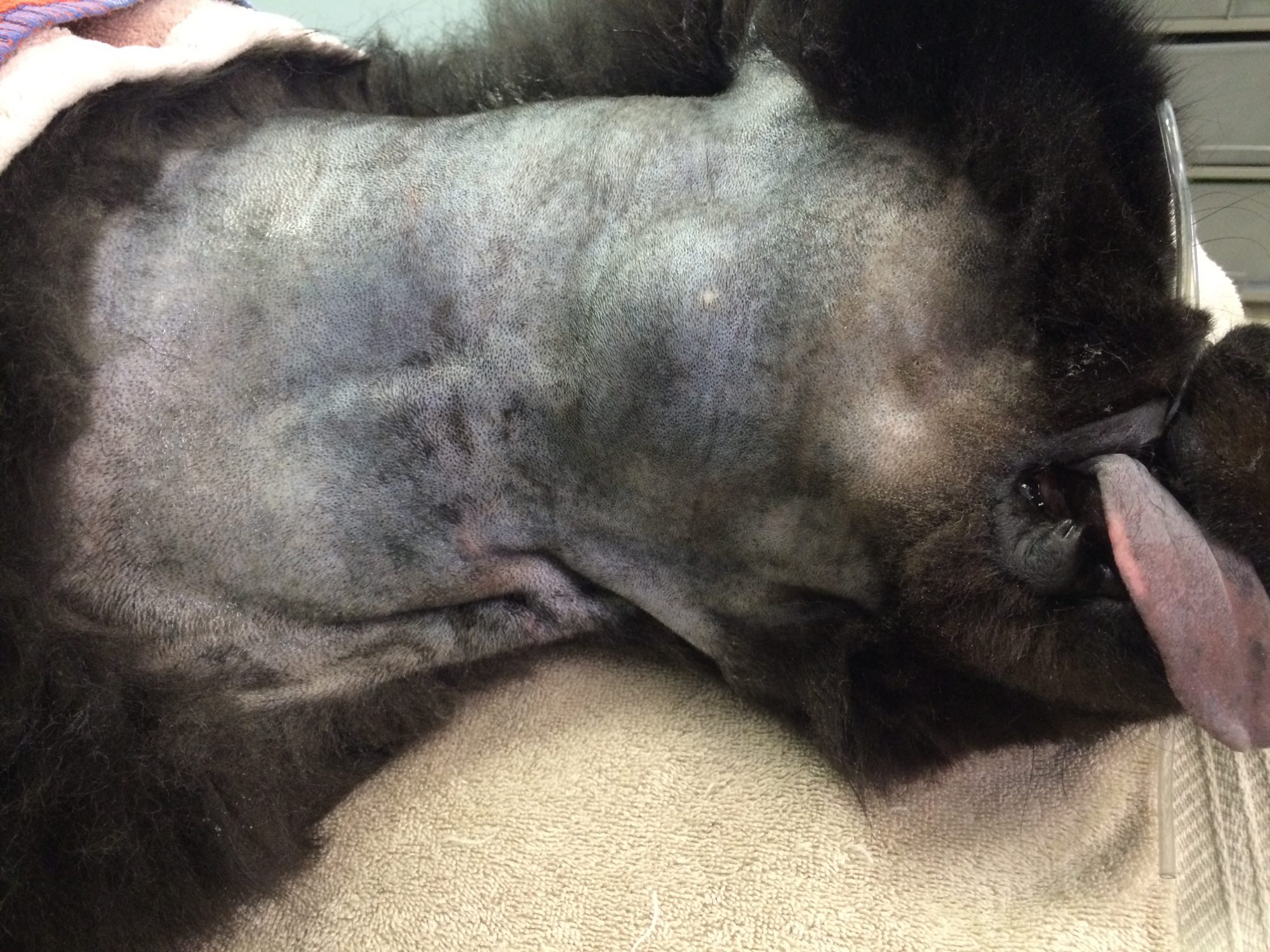 Can you spot the swelling? |
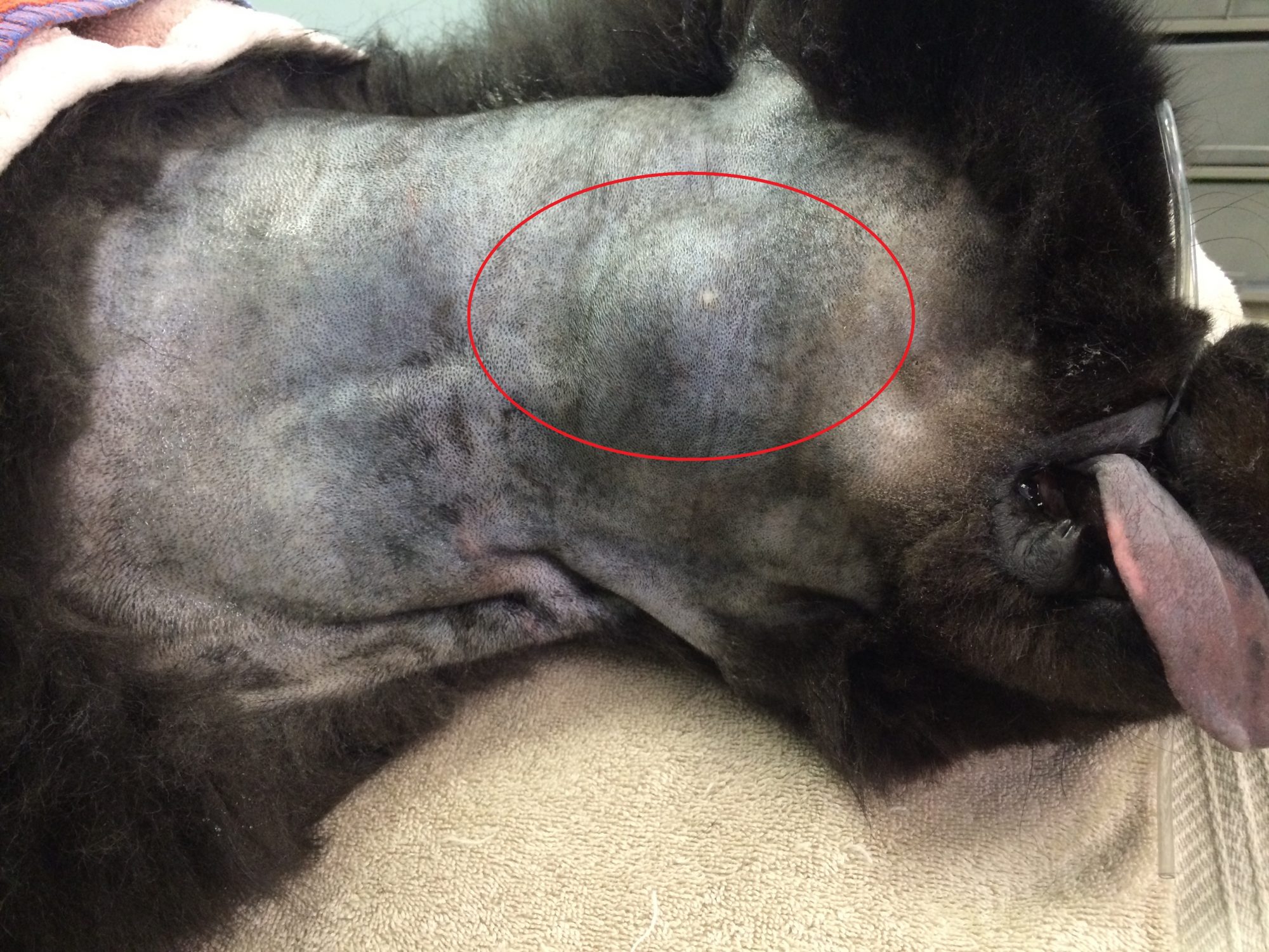 There area within the red circle is the problem area. |
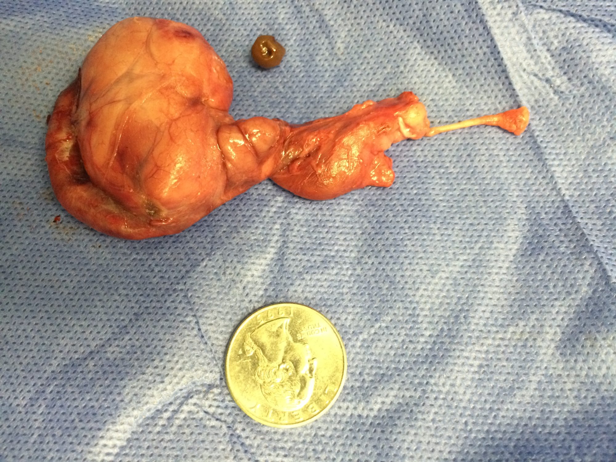 The mass was three times larger initially, but reduced in size after the saliva and several smaller brown masses (also pictured) were removed. |
Dr. Phil Zeltzman is a traveling veterinary surgeon in Pennsylvania & New Jersey. An award-winning author, he loves to share his adventures in practice along with information about vet medicine and surgery that can really help your pets. Dr. Zeltzman specializes in orthopedic, neurologic, cancer, and soft tissue surgeries for dogs, cats, and small exotics. By working with local family vets, he offers the best surgical care, safest anesthesia, and utmost pain management to all his patients. Sign up to get an email when he updates his blog, and follow him on Facebook, too!
Labrador with laryngeal paralysis hits the road to recovery
They say that a journey begins with a single step, but sometimes that step is into your car so you can drive five hours for a scheduled surgery!
Hershey’s owner drove all the way from Jamestown, New York near Lake Erie so I could perform surgery at Barton Heights Veterinary Hospital in Pennsylvania. It was quite the road trip.
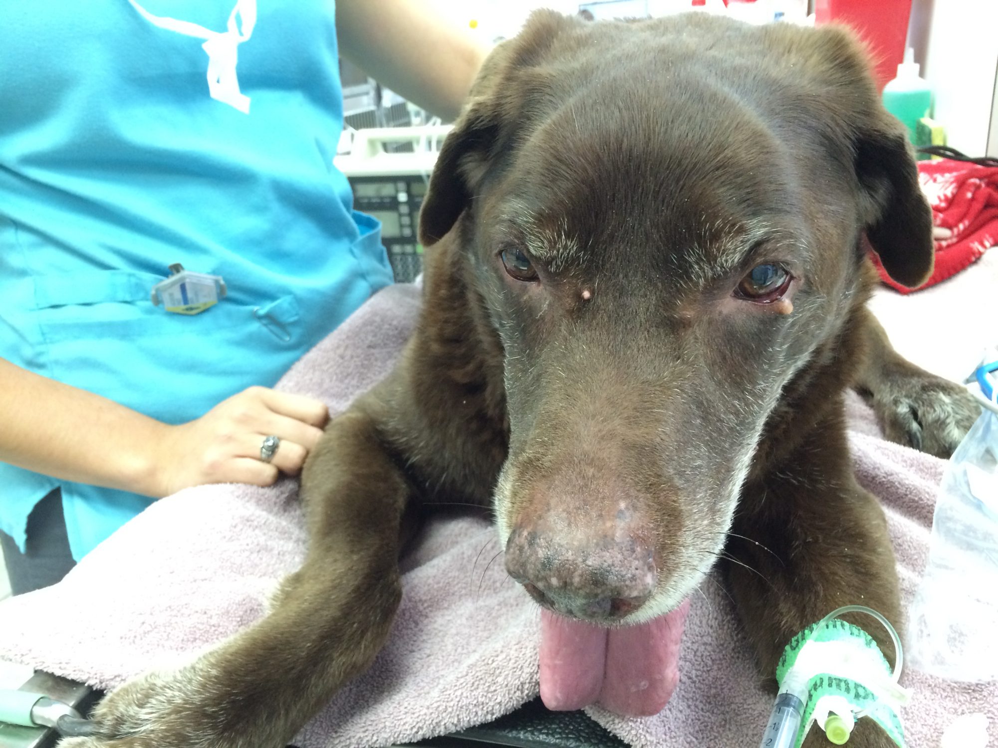
Hershey is an eleven-year-old Lab whose laryngeal paralysis made it difficult for him to breathe. A complicating factor is that he’s diabetic. Hershey’s vet was not sure that surgery was the best bet, but his owner loves Hershey and wasn’t ready to give up on her best friend.
You can see a video of Hershey and his surgery below. He struggles to breathe before the surgery, but after we perform a “tie back” procedure there’s a nice, wide opening. This lets oxygen get in. A tie back surgery uses nylon sutures to keep one side of the larynx open. Two hours after surgery, you can hear the difference! There’s no more struggling and just nice quiet breathing.
You can also see that a small tumor on Hershey’s eyelid was removed. That is a benign tumor called a chalazion, or an adenoma of a Meibomian gland.
Hershey spent a restful night at Barton Heights, and left the hospital for a five hour drive home!
Dr. Phil Zeltzman is a traveling veterinary surgeon in Pennsylvania & New Jersey. An award-winning author, he loves to share his adventures in practice along with information about vet medicine and surgery that can really help your pets. Dr. Zeltzman specializes in orthopedic, neurologic, cancer, and soft tissue surgeries for dogs, cats, and small exotics. By working with local family vets, he offers the best surgical care, safest anesthesia, and utmost pain management to all his patients. Sign up to get an email when he updates his blog, and follow him on Facebook, too!